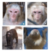
Spontaneous Vitiligo in a Captive Rhesus Monkey (Macaca Mulatta)
- PMID: 37263754
- PMCID: PMC10290481
- DOI: 10.30802/AALAS-CM-22-000091
Spontaneous Vitiligo in a Captive Rhesus Monkey (Macaca Mulatta)
Abstract
Vitiligo affects a significant portion of human and animal populations. The disease causes irregular and multifocal progressive loss of fur, skin, and mucous membrane pigmentation due to the loss or absence of melanocytes. While etiopathogenesis is not completely understood, autoimmunity, environmental, and genetic factors are implicated We present a case report on a 16-y-old female rhesus macaque (Macaca mulatta ) with depigmented areas that are progressively increasing on the skin and coat and are distributed on the head and back. Histopathology revealed alterations compatible with vitiligo characterized by the absence of melanocytes in the epidermis and dermis. The clinical history and complementary exams support this diagnosis.
Figures


Similar articles
-
Pentachrome vitiligo.J Am Acad Dermatol. 1995 Nov;33(5 Pt 2):853-6. doi: 10.1016/0190-9622(95)90422-0. J Am Acad Dermatol. 1995. PMID: 7593796
-
Water Buffalo (Bubalus bubalis) as a spontaneous animal model of Vitiligo.Pigment Cell Melanoma Res. 2016 Jul;29(4):465-9. doi: 10.1111/pcmr.12485. Pigment Cell Melanoma Res. 2016. PMID: 27124831
-
Immunolocalization of tenascin-C in vitiligo.Appl Immunohistochem Mol Morphol. 2012 Oct;20(5):501-11. doi: 10.1097/PAI.0b013e318246c793. Appl Immunohistochem Mol Morphol. 2012. PMID: 22495383
-
The genetics of vitiligo.J Invest Dermatol. 2011 Nov 17;131(E1):E18-20. doi: 10.1038/skinbio.2011.7. J Invest Dermatol. 2011. PMID: 22094401 Free PMC article. Review. No abstract available.
-
Melanocyte-specific, cytotoxic T cell responses in vitiligo: the effective variant of melanoma immunity?Pigment Cell Res. 2005 Aug;18(4):234-42. doi: 10.1111/j.1600-0749.2005.00244.x. Pigment Cell Res. 2005. PMID: 16029417 Review.
References
-
- Alhaidari Z. 2000. Vitiligo chez un chat. Ann Dermatol Venereol 127:413. - PubMed
-
- Alikhan A, Felsten LM, Daly M, Petronic-Rosic V. 2011. Vitiligo: A comprehensive overview Part I. Introduction, epidemiology, quality of life, diagnosis, differential diagnosis, associations, histopathology, etiology, and work-up. J Am Acad Dermatol 65:473e91. 10.1016/j.jaad.2010.11.061 - DOI - PubMed
-
- Bradley BJ, Gerald MS, Widdig A, Mundy NI. 2013. Coat color variation and pigmentation gene expression in rhesus macaques (Macaca mulatta). J Mamm Evol 20:263–270. 10.1007/s10914-012-9212-3. - DOI
Publication types
MeSH terms
LinkOut - more resources
Full Text Sources
Medical

