

Structural interplay of anesthetics and paralytics on muscle nicotinic receptors
- PMID: 37264005
- PMCID: PMC10235084
- DOI: 10.1038/s41467-023-38827-5
Structural interplay of anesthetics and paralytics on muscle nicotinic receptors
Abstract
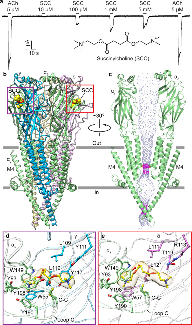
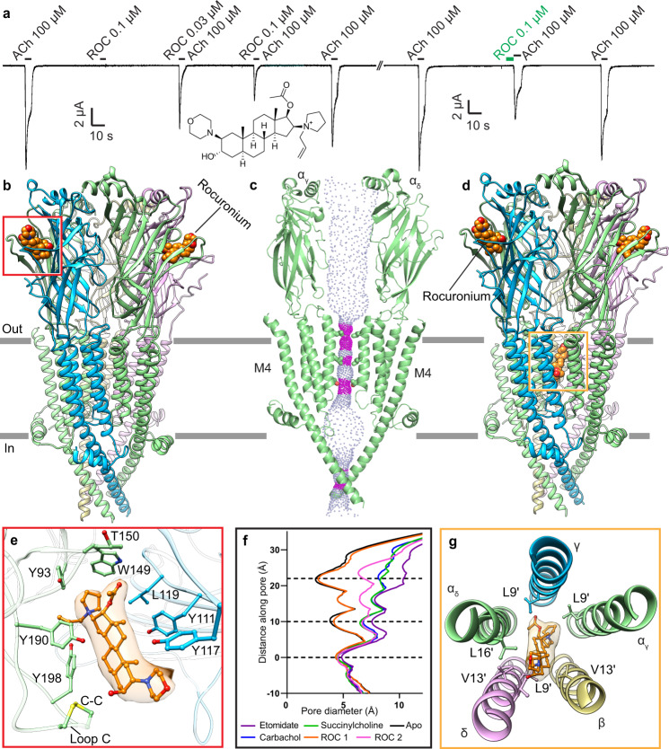
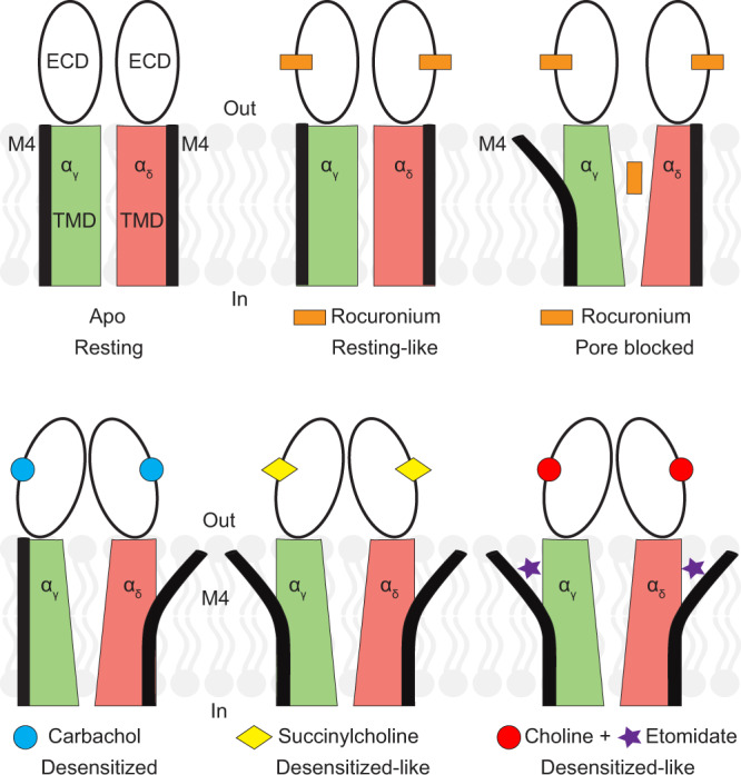
General anesthetics and neuromuscular blockers are used together during surgery to stabilize patients in an unconscious state. Anesthetics act mainly by potentiating inhibitory ion channels and inhibiting excitatory ion channels, with the net effect of dampening nervous system excitability. Neuromuscular blockers act by antagonizing nicotinic acetylcholine receptors at the motor endplate; these excitatory ligand-gated ion channels are also inhibited by general anesthetics. The mechanisms by which anesthetics and neuromuscular blockers inhibit nicotinic receptors are poorly understood but underlie safe and effective surgeries. Here we took a direct structural approach to define how a commonly used anesthetic and two neuromuscular blockers act on a muscle-type nicotinic receptor. We discover that the intravenous anesthetic etomidate binds at an intrasubunit site in the transmembrane domain and stabilizes a non-conducting, desensitized-like state of the channel. The depolarizing neuromuscular blocker succinylcholine also stabilizes a desensitized channel but does so through binding to the classical neurotransmitter site. Rocuronium binds in this same neurotransmitter site but locks the receptor in a resting, non-conducting state. Together, this study reveals a structural mechanism for how general anesthetics work on excitatory nicotinic receptors and further rationalizes clinical observations in how general anesthetics and neuromuscular blockers interact.
© 2023. The Author(s).
Conflict of interest statement
The authors declare no competing interests.
Figures

Similar articles
-
Anesthetic sites and allosteric mechanisms of action on Cys-loop ligand-gated ion channels.Can J Anaesth. 2011 Feb;58(2):191-205. doi: 10.1007/s12630-010-9419-9. Epub 2011 Jan 7. Can J Anaesth. 2011. PMID: 21213095 Free PMC article. Review.
-
Multiple transmembrane binding sites for p-trifluoromethyldiazirinyl-etomidate, a photoreactive Torpedo nicotinic acetylcholine receptor allosteric inhibitor.J Biol Chem. 2011 Jun 10;286(23):20466-77. doi: 10.1074/jbc.M111.219071. Epub 2011 Apr 15. J Biol Chem. 2011. PMID: 21498509 Free PMC article.
-
Synthesis of trifluoromethylaryl diazirine and benzophenone derivatives of etomidate that are potent general anesthetics and effective photolabels for probing sites on ligand-gated ion channels.J Med Chem. 2006 Aug 10;49(16):4818-25. doi: 10.1021/jm051207b. J Med Chem. 2006. PMID: 16884293
-
Intravenous anesthetics differentially modulate ligand-gated ion channels.Anesthesiology. 2000 May;92(5):1418-25. doi: 10.1097/00000542-200005000-00033. Anesthesiology. 2000. PMID: 10781289
-
The role of nicotinic acetylcholine receptors in the mechanisms of anesthesia.Brain Res Bull. 2002 Jan 15;57(2):133-50. doi: 10.1016/s0361-9230(01)00740-7. Brain Res Bull. 2002. PMID: 11849819 Review.
Cited by
-
Effect of Intravenous Lidocaine on Rocuronium: A Randomized Controlled Trial.Kobe J Med Sci. 2025 Feb 12;70(4):E143-E151. doi: 10.24546/0100493127. Kobe J Med Sci. 2025. PMID: 39993787 Free PMC article. Clinical Trial.
-
Influence of lipid bilayer on the structure of the muscle-type nicotinic acetylcholine receptor.Proc Natl Acad Sci U S A. 2024 May 7;121(19):e2319913121. doi: 10.1073/pnas.2319913121. Epub 2024 Apr 29. Proc Natl Acad Sci U S A. 2024. PMID: 38683987 Free PMC article.
-
Autoimmune mechanisms elucidated through muscle acetylcholine receptor structures.Cell. 2025 May 1;188(9):2390-2406.e20. doi: 10.1016/j.cell.2025.03.004. Epub 2025 Apr 8. Cell. 2025. PMID: 40203823
References
-
- Butterworth Iv, J. F., Mackey, D. C. & Wasnick, J. D. Morgan & Mikhail’s Clinical Anesthesiology 6th edn (McGraw-Hill Education, 2018).
