Review
. 2023 Aug;56(2):301-314.
doi: 10.1007/s11239-023-02834-6. Epub 2023 Jun 1.

Positive antiphospholipid antibodies: observation or treatment?

Affiliations
Review

Positive antiphospholipid antibodies: observation or treatment?

Bogna Grygiel-Górniak et al. J Thromb Thrombolysis. 2023 Aug.

Abstract

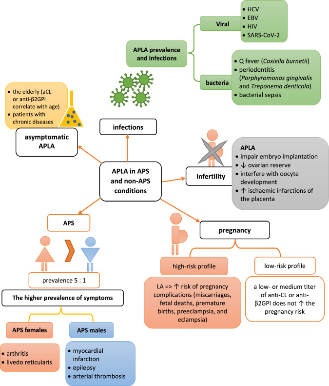
Antiphospholipid antibodies (APLAs) are primarily directed toward phospholipid-binding proteins and are responsible for thrombotic events. APLAs include anti-β2Glycoprotein I (anti-β2GPI), anticardiolipin (anti-CL) antibodies, and lupus anticoagulant. These antibodies are typical markers of antiphospholipid syndrome (APS) and are a part of its diagnostic criteria. Many data underline the presence of APLAs in other rheumatic diseases (e.g., systemic lupus erythematosus, systemic sclerosis, Sjögren's syndrome, rheumatoid arthritis and Behçet's disease). However, they are also detected in patients with cancer, infection, and neurological disorders. Furthermore, healthy individuals may be carriers of APLAs. Chronic asymptomatic APLAs presence is most common in the elderly and subjects with chronic diseases (including malignancies). Specific kinds of APLAs are considered markers of oncological progression. These antibodies occur in 6% of pregnant women (without diagnosed APS) and are related to many pregnancy complications. Of worth, various types of APLAs are reported to have different prothrombotic properties. The risk of thrombotic events in APLA-positive but clinically naïve patients raises many questions in clinical practice. This manuscript analyses various clinical situations and consequences of the APLAs' presence, particularly in patients without diagnosed APS. The prevalence, etiology, molecular background, and prothrombotic properties of numerous APLAs are broadly discussed. The new management approach in different clinical conditions and organ complications is present in the context of recent recommendations. Discussed data underlines that adequate and timely introduced thromboprophylaxis can decrease the risk of thrombus formation and prevent increased morbidity.

Keywords: APLAs in a healthy population; Antiphospholipid antibodies; Molecular mechanisms; Rheumatic and non-rheumatic diseases; Treatment.


Conflict of interest statement

None declared.

Figures

Fig. 1
Fig. 1
The graph presenting literature search methods used in this review
Fig. 2
Fig. 2
The pathological background of APLAs activity. Lupus anticoagulant (LA) binds to phospholipids and does not enable attaching the prothrombinase complex to the cell. The prothrombinase complex (factor Xa and Va) assembles on negatively charged phospholipid membranes in the presence of calcium ions. The prothrombinase complex catalyzes the conversion of prothrombin to thrombin. The enzyme thrombin has procoagulant activity because it converts fibrinogen to fibrin; however, if it binds to thrombomodulin and the endothelial protein C receptor (EPCR), it reveals anticoagulant properties by activating protein C (APC). APC cleaves activated cofactors Va, VIIIa, and plasminogen activator inhibitor (PAI-1), causing a hypocoagulable and hyperfibrinolytic state. Thus, LA is a class of APLAs, which causes a phospholipid-dependent prolongation of the clotting time but is associated with an increased risk of thrombosis and pregnancy complications. Beta 2—glycoprotein I (β2-GPI) consist in five domains (I–V), which can be present in two forms: open (J-shaped) and closed (circular). Domain V (DV) binds phospholipid, and its post-translational modifications cause a conformational change from the circular (closed) form to the open configuration. DV is responsible for binding to cell membranes. The open configuration causes the exposition of a previously hidden domain I (DI) epitope, which becomes a place of antibody binding. Anticardiolipin antibodies (anti-aCL) bind to cardiolipin on the mitochondrial surface and stimulate inflammation. Exacerbated inflammatory processes activate coagulation cascade and thrombosis
Fig. 3
Fig. 3
The prevalence and gender-related characteristics of APS
Fig. 4
Fig. 4
Indications for APLA measurement. VTE venous thromboembolism

References

    1. Tektonidou MG, Andreoli L, Limper M, et al. EULAR recommendations for the management of antiphospholipid syndrome in adults. Ann Rheum Dis. 2019;78:1296–1304. doi: 10.1136/annrheumdis-2019-215213. - DOI - PMC - PubMed
    1. Sammaritano LR. Antiphospholipid syndrome. Best Pract Res Clin Rheumatol. 2020;34:101463. doi: 10.1016/j.berh.2019.101463. - DOI - PubMed
    1. Girón-González JA, García del Río E, Rodríguez C, et al. Antiphospholipid syndrome and asymptomatic carriers of antiphospholipid antibody: prospective analysis of 404 individuals. J Rheumatol. 2004;31:1560–1567. - PubMed
    1. Gezer S. Antiphospholipid syndrome. Dis–Mon DM. 2003;49:696–741. doi: 10.1016/j.disamonth.2003.10.001. - DOI - PubMed
    1. Sciascia S, Baldovino S, Schreiber K, et al. Thrombotic risk assessment in antiphospholipid syndrome: the role of new antibody specificities and thrombin generation assay. Clin Mol Allergy CMA. 2016;14:6. doi: 10.1186/s12948-016-0043-2. - DOI - PMC - PubMed

Substances