Review
. 2023 Jun 1;27(1):55.
doi: 10.1186/s40824-023-00393-8.

Trends in mechanobiology guided tissue engineering and tools to study cell-substrate interactions: a brief review

Affiliations
Review

Trends in mechanobiology guided tissue engineering and tools to study cell-substrate interactions: a brief review

Arun Kumar Rajendran et al. Biomater Res. .

Abstract

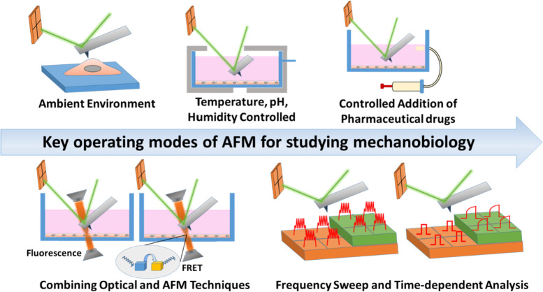
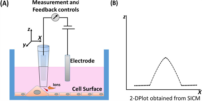
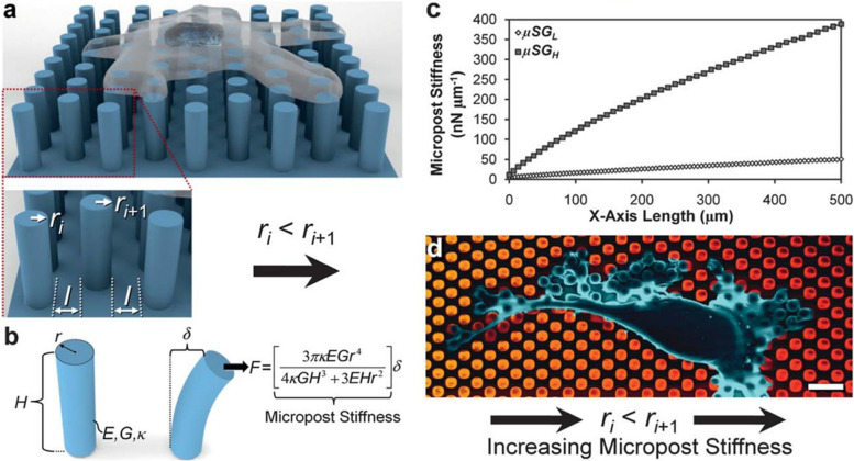
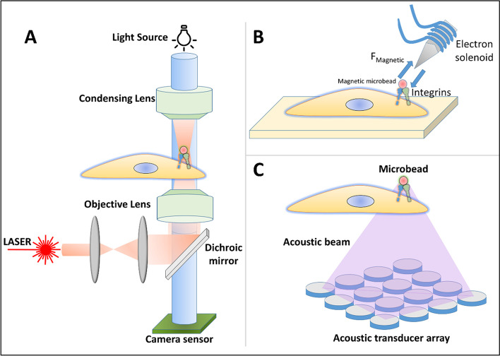
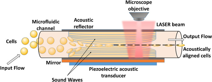
Sensing the mechanical properties of the substrates or the matrix by the cells and the tissues, the subsequent downstream responses at the cellular, nuclear and epigenetic levels and the outcomes are beginning to get unraveled more recently. There have been various instances where researchers have established the underlying connection between the cellular mechanosignalling pathways and cellular physiology, cellular differentiation, and also tissue pathology. It has been now accepted that mechanosignalling, alone or in combination with classical pathways, could play a significant role in fate determination, development, and organization of cells and tissues. Furthermore, as mechanobiology is gaining traction, so do the various techniques to ponder and gain insights into the still unraveled pathways. This review would briefly discuss some of the interesting works wherein it has been shown that specific alteration of the mechanical properties of the substrates would lead to fate determination of stem cells into various differentiated cells such as osteoblasts, adipocytes, tenocytes, cardiomyocytes, and neurons, and how these properties are being utilized for the development of organoids. This review would also cover various techniques that have been developed and employed to explore the effects of mechanosignalling, including imaging of mechanosensing proteins, atomic force microscopy (AFM), quartz crystal microbalance with dissipation measurements (QCMD), traction force microscopy (TFM), microdevice arrays, Spatio-temporal image analysis, optical tweezer force measurements, mechanoscanning ion conductance microscopy (mSICM), acoustofluidic interferometric device (AID) and so forth. This review would provide insights to the researchers who work on exploiting various mechanical properties of substrates to control the cellular and tissue functions for tissue engineering and regenerative applications, and also will shed light on the advancements of various techniques that could be utilized to unravel the unknown in the field of cellular mechanobiology.

Keywords: Cell differentiation; Cell-substrate interaction; Mechanical cues; Mechanobiology; Mechanobiology tools; Organoids.


Conflict of interest statement

The authors declare no competing interests.

Figures

Fig. 1
Fig. 1
Timeline showing some of most significant events associated with mechanobiology
Fig. 2
Fig. 2
Representative model of substrate mediated osteogenic differentiation wherein substrate stiffness (mechanical signal) activates mechanotransduction pathway involving YAP/TAZ (1 A-1B) and non-mechanical signals from substrate leads to osteogenesis via canonical BMPR signaling pathway (2). Adapted with permission from [68]
Fig. 3
Fig. 3
Representative schematic showing tenogenesis in MSCs mediated by mechanotransduction. A combinatorial effect of topography and surface functionalization induced tenogenesis via rhoA activation. Adapted with permission from [88]
Fig. 4
Fig. 4
Representative mechanism of neurogenesis via mechanotransduction wherein the nanotopography manipulates the focal adhesion signaling pathway and neurogenic differentiation. Adapted with permission from [113]
Fig. 5
Fig. 5
Schematic representation of bio-physical forces (stiffness, geometry, fluid shear stress & compression) that could control the formation of organoids. The image was partly created with BioRender.com
Fig. 6
Fig. 6
Schematic representation of traction force in a single cell (A), an overview of cell-cultured on PA gel with fluorescently labeled beads with cells generating traction force with bead displacement (B & C). The image was reproduced with permission from Elsevier [147]
Fig. 7
Fig. 7
Various key operating modes of AFM and its complementary techniques for studying mechanobiology, including (1) bio-imaging: studying mechanical properties in an aqueous environment, (2) studying in the customized chamber for controlling temperature, pH, and humidity with complimentary light microscope imaging, (3) studying the mechanical properties with the addition of pharmaceutical agents, (4) combining optical microscopy techniques and AFM for simultaneous studying of mechanical and biological properties and (5) frequency sweep and time-dependent analysis of mechanical properties of cells
Fig. 8
Fig. 8
A schematic showing the basic assembly of Scanning Ion Conductance Microscope (SICM) (A); A 2-D plot profile that could be obtained after scanning the surface of cells (B)
Fig. 9
Fig. 9
Schematic representation of QCM-D sensor with piezoelectric AT-cut quartz crystal (for using between 0.5 MHz to 300 MHz) having gold electrodes (A) and schematic representation of working of QCM-D with a recording of Δf and ΔD (B). Reproduced under Creative Commons CC BY from [184]
Fig. 10
Fig. 10
Illustration showing the micropillar array which are being utilized for studying mechanobiology. Cell attached over a micropillar surface (a); Pillars showing the deflection due to varied stiffness and the force exerted by the cells (b); the tunability of the micropillar deflection according to the need by changing the stiffness (c); False colored SEM image of attached cell over a gradient micropost array increasing in stiffness (d). Reproduced with kind permission from RSC publishing [194]
Fig. 11
Fig. 11
Representative schematics showing various types of tweezers available for mechanical probing of cells. Optical trap/tweezers (A); Electromagnetic tweezers (B); Acoustic tweezers (C)
Fig. 12
Fig. 12
Basic schematic of Acousto fluidic Interferometric Device (AID). Acoustic reflectors could be lined on both sides of the microfluidic channel, further lined by mirror on bottom side and placed in close contact with the acoustic transducers such as piezoelectric arrays. The acoustic wave from the transducer forms an acoustic plane, thereby aiding in the mechanical alignment of the inflowing cells. When coupled with an interferometer, fringe patterns could be observed and from this data, the physical properties of cells could be interpreted

References

    1. Bear JE, Krause M, Gertler FB. Regulating cellular actin assembly. Curr Opin Cell Biol. 2001;13(2):158–66. doi: 10.1016/S0955-0674(00)00193-9. - DOI - PubMed
    1. Kurosawa M. Phosphorylation and dephosphorylation of protein in regulating cellular function. J Pharmacol Toxicol Methods. 1994;31(3):135–9. doi: 10.1016/1056-8719(94)90075-2. - DOI - PubMed
    1. Berven LA, Crouch MF. Cellular function of p70S6K: a role in regulating cell motility. Immunol Cell Biol. 2000;78(4):447–51. doi: 10.1046/j.1440-1711.2000.00928.x. - DOI - PubMed
    1. Werb Z. ECM and cell surface proteolysis: regulating cellular ecology. Cell. 1997;91(4):439–42. doi: 10.1016/S0092-8674(00)80429-8. - DOI - PubMed
    1. Wells RG. The role of matrix stiffness in regulating cell behavior. Hepatology. 2008;47(4):1394–400. doi: 10.1002/hep.22193. - DOI - PubMed