

Base MRI Imaging Characteristics of Meningioma Patients to Discuss the WHO Classification of Brain Invasion Otherwise Benign Meningiomas
- PMID: 37264676
- PMCID: PMC10272700
- DOI: 10.1177/15330338231171470
Base MRI Imaging Characteristics of Meningioma Patients to Discuss the WHO Classification of Brain Invasion Otherwise Benign Meningiomas
Abstract
Purpose: Compared and analyzed the MRI imaging features of brain invasion otherwise benign (BIOB) meningiomas and WHO grade 1, grade 2 meningiomas, discussed the WHO grading of BIOB from the perspective of imaging.
Materials and methods: A retrospective analysis was performed on 675 meningiomas patients who carried on MRI examination from January 2006 to February 2022. Setting the 2022 Central nervous system (CNS) WHO Guidelines as the gold standard for pathological diagnosis. Statistical analysis of age, gender, and MRI features of meningiomas in relation to WHO grade and brain invasion.
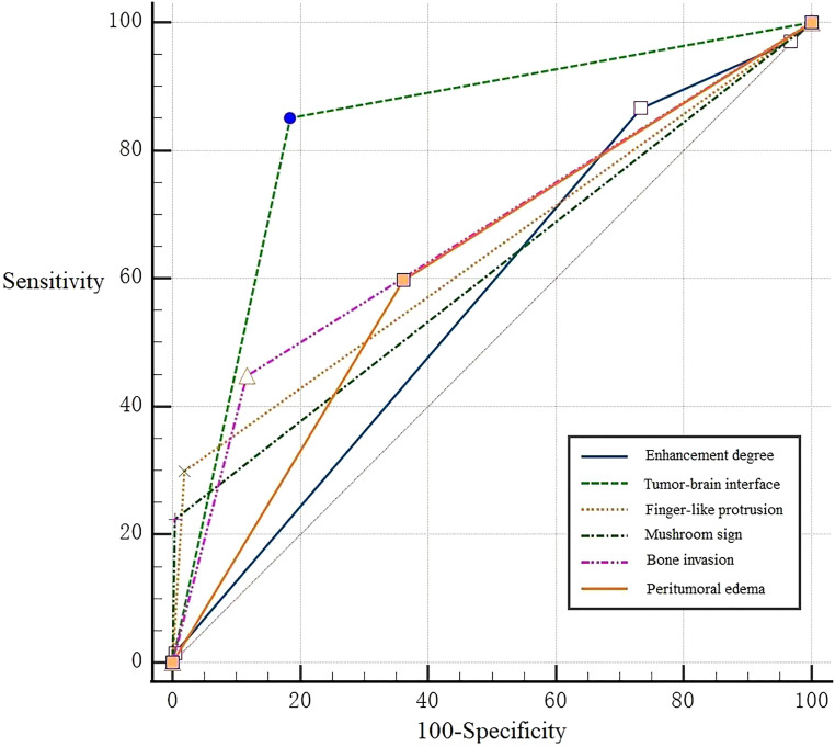
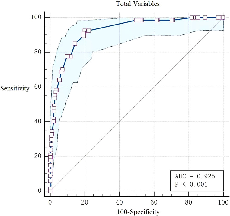
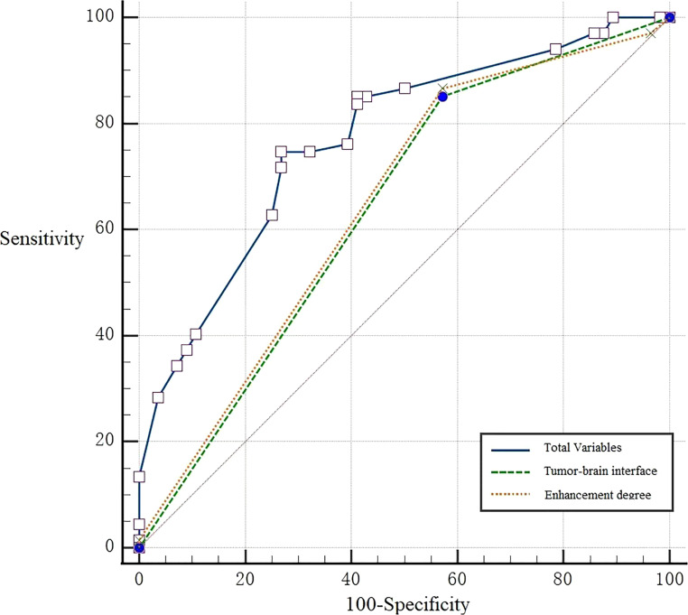
Results: Among 675 cases meningiomas, 543 (80.4%) were WHO grade 1, 123 (18.2%) were WHO grade 2, and 9 (1.3%) were WHO grade 3. There were 108 cases meningiomas with brain invasion (BI) (16.0%) and 567 cases without BI (84.0%). Among BI cases, 67 cases were BIOB. Compared the MRI features between BIOB and WHO grade 1 meningiomas, multivariate analysis demonstrated that the most strongly factors associated with distinguish them were enhancement degree, peritumoral edema, tumor-brain interface, fingerlike protrusion, mushroom sign, and bone invasion (AUC: 0.925 (0.901∼0.945), sensitivity: 0.925, specificity: 0.801). Compared the MRI features between BIOB and WHO grade 2 meningiomas, multivariate analysis demonstrated that the most strongly factors associated with distinguish them were enhancement degree and the tumor-brain interface (AUC: 0.779 (0.686∼0.841), sensitivity: 0.746, specificity: 0.732), their efficacy was slightly weaker.
Conclusions: BIOB is more similar to WHO grade 2 meningiomas in clinical and imaging features than WHO grade 1, so we think that it may be reasonable to classify BIOB as WHO Grade 2 meningiomas in the guidelines.
Keywords: BIOB; Brain invasion; Magnetic Resonance Imaging (MRI); WHO grade.
Figures
Similar articles
-
A radiopathological classification of dural tail sign of meningiomas.J Neurosurg. 2012 Oct;117(4):645-53. doi: 10.3171/2012.6.JNS111987. Epub 2012 Jul 27. J Neurosurg. 2012. PMID: 22839654
-
MRI predictors for brain invasion in meningiomas.Neuroradiol J. 2021 Feb;34(1):3-7. doi: 10.1177/1971400920953417. Epub 2020 Sep 14. Neuroradiol J. 2021. PMID: 32924772 Free PMC article.
-
Dynamic susceptibility contrast and dynamic contrast-enhanced MRI characteristics to distinguish microcystic meningiomas from traditional Grade I meningiomas and high-grade gliomas.J Neurosurg. 2017 Apr;126(4):1220-1226. doi: 10.3171/2016.3.JNS14243. Epub 2016 Jun 10. J Neurosurg. 2017. PMID: 27285539
-
Prognostic significance of brain invasion in meningiomas: systematic review and meta-analysis.Brain Tumor Pathol. 2021 Apr;38(2):81-95. doi: 10.1007/s10014-020-00390-y. Epub 2021 Jan 6. Brain Tumor Pathol. 2021. PMID: 33403457
-
Meningioma grading via diagnostic imaging: A systematic review and meta-analysis.Neuroradiology. 2024 Aug;66(8):1301-1310. doi: 10.1007/s00234-024-03404-0. Epub 2024 Jun 21. Neuroradiology. 2024. PMID: 38902484 Free PMC article.
Cited by
-
Meningiomas with CNS invasion.Front Neurosci. 2023 Jun 29;17:1189606. doi: 10.3389/fnins.2023.1189606. eCollection 2023. Front Neurosci. 2023. PMID: 37456997 Free PMC article. Review.
References
-
- Euskirchen P, Peyre M. Management of meningioma. Presse Med. 2018;47(11-12 Pt 2):e245-e252. - PubMed
-
- Nakasu S, Nakasu Y. Prognostic significance of brain invasion in meningiomas: Systematic review and meta-analysis. Brain Tumor Pathol. 2021;38(2):81-95. - PubMed
-
- Perry JR, Tucker WS, Chui M, Bilbao JM. Dural cavernous hemangioma: An under-recognized lesion mimicking meningioma. Can J Neurol Sci. 1993;20(3):230-233. - PubMed
-
- Perry A, Stafford SL, Scheithauer BW, Suman VJ, Lohse CM. Meningioma grading: An analysis of histologic parameters. Am J Surg Pathol. 1997;21(12):1455-1465. - PubMed
Publication types
MeSH terms
LinkOut - more resources
Full Text Sources
