. 2023 May 1;16(5):dmm049488.
doi: 10.1242/dmm.049488. Epub 2023 Jun 2.

Deficiency in hereditary hemorrhagic telangiectasia-associated Endoglin elicits hypoxia-driven heart failure in zebrafish

Affiliations

Deficiency in hereditary hemorrhagic telangiectasia-associated Endoglin elicits hypoxia-driven heart failure in zebrafish

Etienne Lelièvre et al. Dis Model Mech. .

Abstract

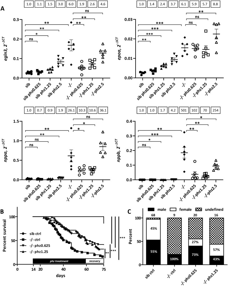
Hereditary hemorrhagic telangiectasia (HHT) is a rare genetic disease caused by mutations affecting components of bone morphogenetic protein (BMP)/transforming growth factor-β (TGF-β) signaling in endothelial cells. This disorder is characterized by arteriovenous malformations that are prone to rupture, and the ensuing hemorrhages are responsible for iron-deficiency anemia. Along with activin receptor-like kinase (ALK1), mutations in endoglin are associated with the vast majority of HHT cases. In this study, we characterized the zebrafish endoglin locus and demonstrated that it produces two phylogenetically conserved protein isoforms. Functional analysis of a CRISPR/Cas9 zebrafish endoglin mutant revealed that Endoglin deficiency is lethal during the course from juvenile stage to adulthood. Endoglin-deficient zebrafish develop cardiomegaly, resulting in heart failure and hypochromic anemia, which both stem from chronic hypoxia. endoglin mutant zebrafish display structural alterations of the developing gills and underlying vascular network that coincide with hypoxia. Finally, phenylhydrazine treatment demonstrated that lowering hematocrit/blood viscosity alleviates heart failure and enhances the survival of Endoglin-deficient fish. Overall, our data link Endoglin deficiency to heart failure and establish zebrafish as a valuable HHT model.

Keywords: Cardiomegaly; Endoglin; Endothelial cells; HHT; Heart failure; Hypoxia.


Conflict of interest statement

Competing interests The authors declare no competing or financial interests.

Figures

Fig. 1.
Fig. 1.
Characterization of zebrafish endoglin chromosomal organization, transcripts and expression pattern. (A) Zebrafish endoglin gene exon-intron structure on chromosome 5. Coding exons (black) and non-coding exons (gray) are shown. (B) RT-PCR analysis of endoglin variants. Endoglin long (arrowheads) and short (asterisks) isoform messenger expression in embryo and adult tissue (brain). (C) Protein alignment of zebrafish Endoglin isoforms. Alternative amino acids (aa) of Endoglin short isoform are in bold. Transmembrane aa are in red. (D) Alignment of human, mouse and zebrafish Endoglin short isoform C-terminal region. Identical (black) and similar (gray) aa are shown. TM, transmembrane. (E) RT-PCR analysis of endoglin variants during zebrafish development. Endoglin short isoform (asterisk) and Endoglin long isoform (arrowhead) are shown. hpf, h postfertilization. (F) Left: whole-mount in situ hybridization using endoglin antisense riboprobe on 24 hpf wild-type embryo. Inset: close-up of the trunk region showing differential endoglin expression between the dorsal aorta (DA) and the posterior cardinal vein (PCV). Arrowheads indicate intersegmental vessels. Right: close-up of the head region showing mostly venous endoglin expression. ACeV, anterior cerebral vein; MCeV, middle cerebral vein; PHBC, primordial hindbrain channel; PMBC, primordial midbrain channel; OV, optic vein. Scale bars: 100 µm.
Fig. 2.
Fig. 2.
Endoglin deficiency results in congestive heart failure in zebrafish. (A) Sanger sequencing chromatograms of gRNA-targeted region on endoglin exon 2 in wild-type (eng+/+), heterozygous (eng+/−) and homozygous (eng−/−) mutant zebrafish. Complementary gRNA sequence is indicated below chromatograms. (B) Imaging of 72 hpf sibling and eng−/− zebrafish blood vessel perfusion. Analysis performed in Tg(kdrl:GFP), Tg(gata1:mRFP) background to highlight blood vessels and erythrocytes, respectively. Images are representative of data from siblings (n=10) and eng−/− fish (n=8). Scale bars: 500 µm. (C) Kaplan–Meier representation of eng+/+, eng+/− and eng−/− fish survival. +/+ versus −/− and +/− versus −/−, ***P<0.0001; +/+ versus +/−, not significant (ns) [log-rank (Mantel–Cox) test]. +/+, n=45; +/−, n=104; −/−, n=56. Arrows point to symptomatic siblings later identified as eng+/−. (D) Influence of genotype over sex ratio in individuals aged 3 months and above. Total number of individuals analyzed is indicated above bars. (E) Representative morphology of 30 dpf sibling and eng−/− fish. Note the enlarged cardiac area and overall paleness in eng−/− fish. Scale bars: 1 mm. (F) H&E-stained heart histological sections reveal enlargement and structural alteration of the ventricle, hypochromic red blood cells and swollen surrounding tissue. a, atrium; ba, bulbus arteriosus; v, ventricle. Scale bars: 100 µm.
Fig. 3.
Fig. 3.
Cardiac stress, ventricle enlargement and increased cardiomyocyte number correlate with hypoxia in Endoglin-deficient fish. (A,B) RT-qPCR analysis of nppa and nppb (cardiac stress) (A), and egln3 and epoa (hypoxia) (B), in 5, 10, 15, 20, 25 and 30 dpf sibling and eng−/− fish (RT-PCR genotyping is presented below graphs). Target gene expression values were normalized to rpl13a. 5 dpf and 10 dpf, n=15; 15 dpf, n=8; 20 dpf, n=5; 25 dpf, n=4; 30 dpf, n=3. Data are mean±s.e.m. of technical replicates. nppa: 15 dpf, 20 dpf, 25 dpf and 30 dpf, *P=0.05. nppb: 15 dpf, *P=0.05; 20 dpf, *P=0.0383; 25 dpf and 30 dpf, *P=0.05. egln3: 15 dpf and 20 dpf, *P=0.05; 25 dpf, *P=0.03831; 30 dpf, *P=0.05. epoa: 15 dpf, 20 dpf, 25 dpf and 30 dpf, *P=0.05. ns, not significant (one-tailed Mann–Whitney test). dfp, days postfertilization. (C) Analysis of ventricle volume in 10, 12 and 15 dpf sibling and eng−/− fish in Tg(cmlc2:GFP) background (n=5). Ventricle volume is estimated by the simplified ellipsoid volume calculation formula V=0.523(width in mm)2(length in mm). *P=0.0159; ns, not significant (one-tailed Mann–Whitney test). (D) Flow cytometry analysis of cardiomyocyte (cmlc2:GFP+) fraction in whole organism at 5, 10, 15, 20 and 25 dpf. Cell suspensions at 5, 10, 15, 20 and 25 dpf were prepared from pools of 26, 18, nine, seven and seven eng−/− fish or siblings, respectively. Bottom graph represents fold change in cmlc2:GFP+ cells in eng−/− fish relative to siblings. Data are representative of three independent experiments.
Fig. 4.
Fig. 4.
Abnormal branchial vascular development in Endoglin-deficient fish underlies structural impairment of gills. (A) H&E-stained histological sections of 30 dpf sibling and eng−/− gills. Note the poorly developed lamellae (arrowheads) and enlarged artery (asterisk) on branchial arch 4 (AA6) in eng−/− fish gills. Scale bars: 100 µm. (B) Top three rows: kinetic analysis of gill vascular development in 10, 12 and 15 dpf sibling and eng−/− fish in Tg(kdrl:GFP), Tg(flt1:tdtomato) background. Note the reduced length of afferent filamental artery (GFP+, Tomatohigh) and efferent filamental artery (GFP+, Tomatolow/−) (arrowheads) and enlarged efferent branchial artery) (asterisks) as early as 10 dpf. Note the overall loss of flt1 signal in 15 dpf eng−/− fish. Scale bars: 100 µm. Bottom row: graphical representation of sibling and eng−/− fish branchial efferent artery (EFA) diameter (left) and filamental artery (FA) length (right) at 10, 12 and 15 dpf. EFA diameter and FA length were measured on AA6. EFA diameter was measured at the most dorsal FA level. FAs (five per fish) were measured starting from the most dorsal FA. Data are presented as individual values and mean±s.e.m. EFA diameter: siblings (sib) versus −/−, ***P=0.0003, ***P=0.0006 and ***P=0.0003 at 10, 12 and 15 dpf, respectively. FA length: sib versus −/−, *P=0.037, ***P<0.0001 and **P=0.0026 at 10, 12 and 15 dpf, respectively. One-tailed Mann–Whitney test.
Fig. 5.
Fig. 5.
Phenylhydrazine treatment alleviates pathological conditions induced by Endoglin deficiency in zebrafish. (A) RT-qPCR analysis of egln3, epoa and nppa and nppb expression in 27 dpf sibling and eng−/− fish untreated or treated with 0.625, 1.25 and 2.5 µg/ml phenylhydrazine (phz). Target gene expression is represented as 2−ΔCT using rpl13a as reference. Samples (n=6) are pools of four fish. Data are presented as individual values and mean±s.e.m. Mean fold change using sib as reference is indicated above graphs. egln3: sib versus sib phz 0.625 µg/ml (phz0.625), P=0.1178; sib versus sib phz 1.25 µg/ml (phz1.25), *P=0.0104; sib versus sib phz 2.5 µg/ml (phz2.5), **P=0.0019; sib control (ctrl) versus −/− ctrl, **P=0.0037; −/− ctrl versus −/− phz0.625, **P=0.005; −/− ctrl versus −/− phz1.25, P=0.0103; −/− ctrl versus −/− phz2.5, P=0.0814. epoa: sib versus sib phz0.625, **P=0.0068; sib versus sib phz1.25, ***P<0.0001; sib versus sib phz2.5, ***P=0.0003; sib ctrl versus −/− ctrl, ***P=0.0003; −/− ctrl versus −/− phz0.625, P=0.1573; −/− ctrl versus −/− phz1.25, P=0.1489; −/− ctrl versus −/− phz2.5, **P=0.0094. nppa: sib versus sib phz0.625, P=0.0525; sib versus sib phz1.25, P=0.1850; sib versus sib phz2.5, **P=0.0014; sib ctrl versus −/− ctrl, **P=0.0045; −/− ctrl versus −/− phz0.625, *P=0.0204; −/− ctrl versus −/− phz1.25, *P=0.0206; −/− ctrl versus −/− phz2.5, P=0.0675. nppb: sib versus sib phz0.625, P=0.3146; sib versus sib phz1.25, *P=0.0417; sib versus sib phz2.5, ***P=0.0005; sib ctrl versus −/− ctrl, **P=0.0032; −/− ctrl versus −/− phz0.625, **P=0.0043; −/− ctrl versus −/− phz1.25, **P=0.0042; −/− ctrl versus −/− phz2.5, *P=0.0259. ns, not significant. Shown are false discovery rate (FDR)-adjusted P-values from Brown–Forsythe test and Welch's test, and two-stage linear set-up procedure of Benjamini, Krieger and Yekutieli posthoc test for multiple comparisons after assessment of normal distribution and equal s.d. (B) Phz treatment enhances eng−/− survival. Kaplan–Meier representation of the survival of sibling and eng−/− fish non-treated (ctrl) or treated with phz at 0.625 or 1.25 µg/ml. sib versus −/− ctrl or −/−phz1.25 or −/−phz0.625, ***P<0.0001; −/− versus −/− phz1.25, **P=0.0039; −/− versus −/− phz0.625, ***P=0.0009 [log-rank (Mantel–Cox) test]. sib ctrl, n=66; −/− ctrl, n=55; −/−phz1.25, n=53; −/−phz0.625, n=48. (C) Influence of phz treatment on sex ratio in individuals aged 2.5 months and above. Graph represents the percentage of fish reaching adulthood. Analyzed fish numbers are indicated above bars. Male and female values are indicated inside bars.
Fig. 6.
Fig. 6.
Both long and short Endoglin isoforms rescue eng−/− heart failure. (A) PCR genotyping example of established fli1a:engLong and fli1a:engshort transgenic lines. Amplicon size difference is due to exon 13 inclusion in endoglin short coding transcript. (B) Mutant phenotype frequency in clutches from, eng+/−, Tg(fli1a:engLong) and eng+/−, Tg(fli1a:engshort) with eng+/− fish at 3 dpf. Total number of mutants over total number of fish analyzed is indicated above bars. (C) Genotype distribution frequency and associated survival in 5-month-old fish from crosses between eng+/− and eng+/−, Tg(fli1a:engLong) or eng+/−, Tg(fli1a:engshort). Left column shows even transgene distribution among the different genotypes. Fish numbers are indicated above bars. Right column shows Kaplan–Meier representation of survival according to genotype and transgenic status. −/− versus −/−, Tg(fli1a:engLong) or Tg(fli1a:engshort), ***P<0.0001 [log-rank (Mantel–Cox) test].

References

    1. Arjamaa, O. and Nikinmaa, M. (2011). Hypoxia regulates the natriuretic peptide system. Int. J. Physiol. Pathophysiol. Pharmacol. 3, 191-201. - PMC - PubMed
    1. Baeyens, N., Larrivée, B., Ola, R., Hayward-Piatkowskyi, B., Dubrac, A., Huang, B., Ross, T. D., Coon, B. G., Min, E., Tsarfati, M.et al. (2016). Defective fluid shear stress mechanotransduction mediates hereditary hemorrhagic telangiectasia. J. Cell Biol. 214, 807-816. 10.1083/jcb.201603106 - DOI - PMC - PubMed
    1. Bellón, T., Corbí, A., Lastres, P., Calés, C., Cebrián, M., Vera, S., Cheifetz, S., Massague, J., Letarte, M. and Bernabéu, C. (1993). Identification and expression of two forms of the human transforming growth factor-beta-binding protein endoglin with distinct cytoplasmic regions. Eur. J. Immunol. 23, 2340-2345. 10.1002/eji.1830230943 - DOI - PubMed
    1. Blanco, F. J. and Bernabeu, C. (2011). Alternative splicing factor or splicing factor-2 plays a key role in intron retention of the endoglin gene during endothelial senescence. Aging Cell 10, 896-907. 10.1111/j.1474-9726.2011.00727.x - DOI - PubMed
    1. Blanco, F. J., Grande, M. T., Langa, C., Oujo, B., Velasco, S., Rodriguez-Barbero, A., Perez-Gomez, E., Quintanilla, M., López-Novoa, J. M. and Bernabeu, C. (2008). S-endoglin expression is induced in senescent endothelial cells and contributes to vascular pathology. Circ. Res. 103, 1383-1392. 10.1161/CIRCRESAHA.108.176552 - DOI - PubMed

Publication types

MeSH terms