. 2023 May 17:11:1190630.
doi: 10.3389/fchem.2023.1190630. eCollection 2023.

Improving osteoinduction and osteogenesis of Ti6Al4V alloy porous scaffold by regulating the pore structure

Affiliations

Improving osteoinduction and osteogenesis of Ti6Al4V alloy porous scaffold by regulating the pore structure

Chao Wang et al. Front Chem. .

Abstract

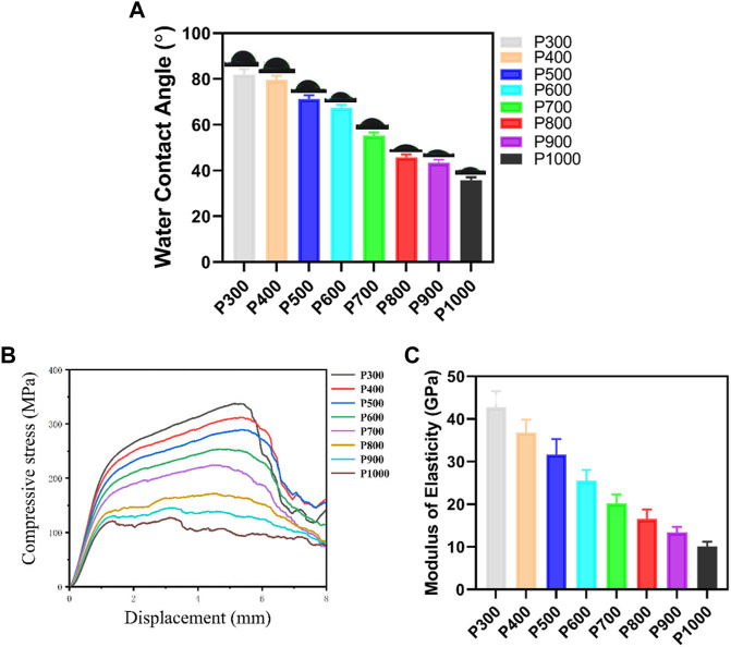
Titanium alloy scaffolds with a porous structure have attracted much attention in bone defect repair. However, which pore structure is more beneficial to bone defect repair is controversial. In the present research, the Ti6Al4V alloy porous scaffolds with gradient pore sizes were designed and fabricated. The microstructure characterization, tests of mechanical properties, and in vitro and in vivo experiments have been performed to systematically evaluate the effect of pore size on osteoinduction and osteogenesis. The results revealed that the contact angle with water, compressive strength, and elastic modulus of the Ti6Al4V alloy porous scaffolds decreased gradually with the increase of pore size. However, there were obvious drops when the pore size of the porous scaffold was around 600 μm. As the pore size increased, the proliferation and integrin β1 of RAW 264.7 macrophages seeded on Ti6Al4V alloy porous scaffolds increased at first, reaching a maximum value at a pore size of around 600 μm, and then decreased subsequently. The proliferation, integrin β1, and osteogenic gene-related expressions of Bone marrow mesenchymal stem cells (BMSCs) seeded on Ti6Al4V alloy porous scaffolds with different pore sizes all exhibited similar variations which rose with increased pore size firstly, obtaining the maximum value at pore size about 600 μm, and then declined. The in vivo experiments confirmed the in vitro results, and the Ti6Al4V alloy porous scaffold with a pore size of 600 μm possessed the better capability to induce new bone formation. Therefore, for the design of Ti6Al4V alloy with a regular porous scaffold, the surface morphology, porosity, strength, and elastic modulus should be considered systematically, which would determine the capability of osteoinduction and osteogenesis.

Keywords: Ti6Al4V alloy; bone defect repairing; osteogenesis; pore structure; porous scaffold.


Conflict of interest statement

The authors declare that the research was conducted in the absence of any commercial or financial relationships that could be construed as a potential conflict of interest.

Figures

FIGURE 1
FIGURE 1
The surgical procedure of porous scaffold implantation in the bone defect. (A) Skin preparation in the operative area to expose the femur. (B) Preparation of the bone defect step by step. (C) Implantation of porous scaffolds with different pore sizes. (D) Suture of the wound.
FIGURE 2
FIGURE 2
Morphology of the Ti6Al4V alloy porous scaffolds and their pore size analysis. (A) Morphology of porous structure by SEM and pore size measured using Image-J software. (B) The macrograph of the porous scaffolds with different pore sizes. (C) Statistical analyses of the pore size in different porous scaffolds.
FIGURE 3
FIGURE 3
Physical and mechanical properties of the Ti6Al4V alloy porous scaffolds with different pore sizes. (A) Variation of contact angle of water on porous scaffolds with different pore sizes. (B) Compressive stress-displacement curves of porous scaffolds with different pore sizes. (C) Elastic modulus of porous scaffolds with different pore sizes.
FIGURE 4
FIGURE 4
The viability and proliferation of macrophages on porous scaffolds with different pore sizes. (A) Detection of macrophage viability on porous scaffolds with different pore sizes. (B) CCK8 test results of macrophage proliferation ability on porous scaffolds with different pore sizes (*p < 0.05, **p < 0.01, and ***p < 0.001).
FIGURE 5
FIGURE 5
Immunofluorescence staining of macrophages on porous scaffolds with different pore sizes (red fluorescence is integrin β1, green fluorescence is cytoskeleton, blue is nucleus).
FIGURE 6
FIGURE 6
Staining image of BMSCs on porous scaffolds with different pore sizes (green fluorescent cells are living cells, red fluorescent cells are dead cells).
FIGURE 7
FIGURE 7
SEM images of BMSCs adhered on porous scaffolds with different pore sizes (yellow arrows point to BMSCs).
FIGURE 8
FIGURE 8
Immunofluorescence staining of BMSCs on porous scaffolds with different pore sizes (red fluorescence is integrin β1, green fluorescence is cytoskeleton, blue is nucleus).
FIGURE 9
FIGURE 9
Immunofluorescence staining images of BMSCs on porous scaffolds with different pore sizes (red fluorescence is integrin β1, green fluorescence is cytoskeleton, blue is nucleus).
FIGURE 10
FIGURE 10
RT-PCR detection on the osteogenic gene-related expression of BMSCs cultured on porous scaffolds with different pore sizes. (*p < 0.05, **p < 0.01, and ***p < 0.001).
FIGURE 11
FIGURE 11
Micro-CT analysis on the porous scaffolds implanted for 12 weeks and the corresponding tissue analyses by H&E and Von Gieson staining. (A) Micro-CT evaluation of bone ingrowth, vertical and transverse images were presented. (B) Quantitative analysis of BV/TV. (*p < 0.05, **p < 0.01, and ***p < 0.001, when compared with P500.) (C) H&E and Von Gieson staining for detecting the new bone formation, trabecular bone ingrowth and integration in each group. (red referring to bone tissue, black referring to the porous scaffold, and the yellow arrow pointing to the new bone along the interface of scaffold and bone tissue).

Similar articles

Cited by

References

    1. Akram F., Kunze K. N., Kerzner B., Gonzalez A., Palacios A., Levine B. R. (2021). Mid- term survivorship, performance, and predictors of outcome in primary total hip arthroplasty with a porous tantalum femoral prosthesis. J. Arthroplasty 36 (5), 1688–1694. 10.1016/j.arth.2020.12.001 - DOI - PubMed
    1. Antonov A. S., Antonova G. N., Munn D. H., Mivechi N., Lucas R., Catravas J. D., et al. (2011). αVβ3 integrin regulates macrophage inflammatory responses via PI3 kinase/Akt-dependent NF-κB activation. J. Cell Physiol. 226 (2), 469–476. 10.1002/jcp.22356 - DOI - PMC - PubMed
    1. Barth H. D., Zimmermann E. A., Schaible E., Tang S. Y., Alliston T., Ritchie R. O. (2011). Characterization of the effects of x-ray irradiation on the hierarchical structure and mechanical properties of human cortical bone. Biomaterials 32 (34), 8892–8904. 10.1016/j.biomaterials.2011.08.013 - DOI - PMC - PubMed
    1. Bassous N. J., Jones C. L., Webster T. J. (2019). 3-D printed Ti-6Al-4V scaffolds for supporting osteoblast and restricting bacterial functions without using drugs: Predictive equations and experiments. Acta Biomater. 96, 662–673. 10.1016/j.actbio.2019.06.055 - DOI - PubMed
    1. Berry D. J., Harmsen W. S., Cabanela M. E., Morrey B. F. (2002). Twenty-five-year survivorship of two thousand consecutive primary charnley total hip replacements: Factors affecting survivorship of acetabular and femoral components. J. Bone Jt. Surg. Am. 84 (2), 171–177. 10.2106/00004623-200202000-00002 - DOI - PubMed