Antenatal diagnosis of hydrometrocolpos with Mullerian duplication on ultrasound and fetal MRI: case report and literature review
- PMID: 37265753
- PMCID: PMC10230225
- DOI: 10.1259/bjrcr.20230024
Antenatal diagnosis of hydrometrocolpos with Mullerian duplication on ultrasound and fetal MRI: case report and literature review
Abstract
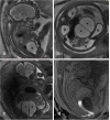
Fetal abdomino-pelvic cystic lesions are uncommon and can have varied etio-pathogenesis. Most commonly they originate from the gastrointestinal or genitourinary tract. These include choledochal cyst, hydronephrosis, renal cyst, mesenteric/omental cyst, ovarian cyst, meconium pseudocyst, and hydrocolpos/hydrometrocolpos among others. Fetal hydrometrocolpos is rare with a reported incidence of 0.006% and its diagnosis requires a high index of suspicion. Antenatal ultrasound and magnetic resonance imaging (MRI) is invaluable in diagnostic evaluation. This case report describes the imaging features of antenatally detected congenital hydrometrocolpos with Mullerian duplication secondary to cloacal malformation using antenatal ultrasound and MRI. Per-operative findings and other possible differential diagnoses are discussed along with a brief review of literature.
© 2023 The Authors. Published by the British Institute of Radiology.
Conflict of interest statement
Competing interests: I hereby declare that this manuscript is original, the authors listed have contributed significantly to the production of this manuscript and that we have not used any sources other than those listed in the bibliography and identified as references. I further declare that we have not submitted this manuscript to any other institution or journal. We declare no conflicts of interest or external funding in the production of this manuscript and have adhered to ethical guidelines.
Figures


References
Publication types
LinkOut - more resources
Full Text Sources

