. 2023 May 23:97:71-116.
doi: 10.3897/mycokeys.97.103118. eCollection 2023.

Botryosphaerialean fungi associated with woody oil plants cultivated in Sichuan Province, China

Affiliations

Botryosphaerialean fungi associated with woody oil plants cultivated in Sichuan Province, China

Wen-Li Li et al. MycoKeys. .

Abstract

Woody oil plants are important economic trees which are widely cultivated and distributed throughout China. Surveys conducted during 2020 and 2021 on several woody oil plantations from five regions of Sichuan Province, China, revealed a high diversity of Botryosphaerialean fungi. The identification of 50 botryosphaeriaceous isolates was carried out based on both morphology and multi-gene phylogenetic analysis of internal transcribed spacer region (ITS), translation elongation factor 1-alpha gene (tef1) and β-tubulin gene (tub2). This allowed the identification of twelve previously known Botryosphaeriales species: Aplosporellaprunicola, A.ginkgonis, Barriopsistectonae, Botryosphaeriadothidea, Bo.fabicerciana, Diplodiamutila, Di.seriata, Dothiorellasarmentorum, Neofusicoccumparvum, Sardiniellaguizhouensis, Sphaeropsiscitrigena, and Sp.guizhouensis, and four novel species belonging to the genera Diplodia and Dothiorella, viz. Di.acerigena, Di.pistaciicola, Do.camelliae and Do.zanthoxyli. The dominant species isolated across the surveyed regions were Botryosphaeriadothidea, Sardiniellaguizhouensis and Diplodiamutila, representing 20%, 14% and 12% of the total isolates, respectively. In addition, most isolates were obtained from Pistaciachinensis (14 isolates), followed by Camelliaoleifera (10 isolates). The present study enhances the understanding of Botryosphaeriales species diversity on woody oil plants in Sichuan Province, China.

Keywords: Botryosphaeriales; diversity; new species; phylogeny; taxonomy.


Figures

Figure 1.
Figure 1.
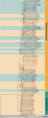
Phylogram generated from RAxML analysis based on combined ITS and tef1 sequence data of Botryosphaeriaceae and Aplosporellaceae isolates. The tree was rooted to Lecanostictaacicula (LNPV 252). The ML (≥ 75%) and BI (≥ 95%) bootstrap supports are given near the nodes, respectively. Isolates from this study are marked in red and ex-type strains are marked in bold.
Figure 2.
Figure 2.
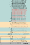
Phylogram generated from RAxML analysis based on combined ITS and tef1 sequence data of Botryosphaeria isolates. The tree was rooted to Barriopsisiraniana (IRAN1448C and IRAN1449C). The ML (≥ 75%) and BI (≥ 95%) bootstrap supports are given near the nodes, respectively. Isolates from this study are marked in red and ex-type strains are marked in bold.
Figure 3.
Figure 3.
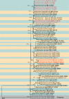
Phylogram generated from RAxML analysis based on combined ITS, tef1 and tub2 sequence data of Diplodia isolates. The tree was rooted to Dothiorelladulcispinae (CMW 36460 and CMW 36462). The ML (≥ 75%) and BI (≥ 95%) bootstrap supports are given near the nodes, respectively. Isolates from this study are marked in red and ex-type strains are marked in bold.
Figure 4.
Figure 4.
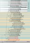
Phylogram generated from RAxML analysis based on combined ITS, tef1 and tub2 sequence data of Dothiorella isolates. The tree was rooted to Neofusicoccumluteum (CBS 562.92 and CMW 41365). The ML (≥ 75%) and BI (≥ 95%) bootstrap supports are given near the nodes, respectively. Isolates from this study are marked in red and ex-type strains are marked in bold.
Figure 5.
Figure 5.
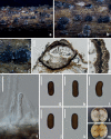
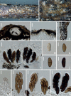
Aplosporellaginkgonis (HUEST 22.0092, new host record) a–c appearance of conidiomata on natural substrate d vertical section of conidioma e section of peridium f conidiogenous cells and developing conidia g–k brown aseptate conidia l upper view of the colony on PDA after 14 d m reverse view of the colony on PDA after 14 d. Scale bars: 100 μm (d); 40 μm (e); 10 μm (f–k).
Figure 6.
Figure 6.
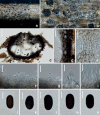
Aplosporellaprunicola (HUEST 22.0091, new host record) a, b appearance of conidiomata on natural substrate c vertical section of multiloculate conidioma d, e section of peridium f–i conidiogenous cells and developing conidia j–n brown aseptate conidia. Scale bars: 100 μm (c); 20 μm (d, e); 10 μm (f–n).
Figure 7.
Figure 7.
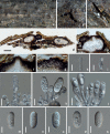
The sexual morph of Diplodiaacerigena (HKAS 125891, holotype) a, b appearance of ascomata on natural substrate c vertical section of ascoma d ostiole e section of peridium f–h asci with hyaline ascospores i asci with brown 2-sepatate ascospores j, k hyaline immature aseptate ascospores l–n mature brown 2-septate ascospores o germinated ascospore p upper view of the colony on PDA after 14 d q reverse view of the colony on PDA after 14 d. Scale bars: 100 μm (c); 10 μm (d, e, j–o); 20 μm (f–i).
Figure 8.
Figure 8.
The asexual morph of Diplodiaacerigena (HKAS 125891, holotype) a–d appearance of conidiomata on PDAe–i conidiogenous cells and developing conidia j, k hyaline immature conidia l–o mature brown aseptate conidia p–t Spermatogenous cells and Spermatia. Scale bars: 10 μm (e–o, t); 5 μm (p–s).
Figure 9.
Figure 9.
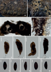
Diplodiamutila (HUEST 22.0069, new host record) a, b appearance of conidiomata on natural substrate c vertical section of conidioma d ostiole e section of peridium f–i conidiogenous cells and developing conidia j hyaline immature conidium k–n mature brown conidia. Scale bars: 40 μm (c); 20 μm (d, e); 10 μm (f–n).
Figure 10.
Figure 10.
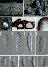
Diplodiapistaciicola (HKAS 125890, holotype) a–c appearance of conidiomata on natural substrate d, e vertical section of conidiomata/conidioma f ostiole g section of peridium h–l conidiogenous cells and developing conidia m–q hyaline aseptate conidia. Scale bars: 200 μm (d); 50 μm (e); 20 μm (f, g); 10 μm (h–q).
Figure 11.
Figure 11.
Diplodiaseriata (HUEST 22.0073, new host record) a–c appearance of ascomata on natural substrate d vertical section of ascomata e section of peridium f–i asci j–n ascospores. Scale bars: 100 μm (d); 10 μm (e, j–n); 20 μm (f–i).
Figure 12.
Figure 12.
Dothiorellacamelliae (HKAS 125892, holotype) a, b appearance of ascomata on natural substrate c vertical section of ascoma d section of peridium e–g asci h–m ascospores n germinated ascospore. Scale bars: 50 μm (c); 10 μm (d, h–n); 20 μm (e–g).
Figure 13.
Figure 13.
Dothiorellasarmentorum (HUEST 22.0077, new host record) a, b appearance of conidiomata on natural substrate c vertical section of conidioma d–g conidiogenous cells and developing conidia h–l brown conidia. Scale bars: 50 μm (c); 10 μm (d–l).
Figure 14.
Figure 14.
Dothiorellazanthoxyli (HKAS 125893, holotype) a, b appearance of ascomata on natural substrate c vertical section of ascoma d ostiole e section of peridium f, k–n asci g–j brown ascospores. Scale bars: 50 μm (c); 20 μm (d–f); 10 μm (g–n).
Figure 15.
Figure 15.
Neofusicoccumparvum (HUEST 22.0097, new host record) a, b appearance of ascomata on natural substrate c, d vertical section of ascomata e ostiole f section of peridium g–j ascospores k, l immature asci m, n mature asci. Scale bars: 100 μm (d); 25 μm (e, f); 10 μm (g–j); 20 μm (k–n).
Figure 16.
Figure 16.
Sardiniellaguizhouensis (HUEST 22.0100, new host record) a–c appearance of conidiomata on natural substrate d vertical section of conidioma e section of peridium f ostiole g–j conidiogenous cells and developing conidia k–n conidia. Scale bars: 40 μm (d); 20 μm (e, f); 10 μm (g–n).
Figure 17.
Figure 17.
Sphaeropsiscitrigena (HUEST 22.0107, new host record) a, b appearance of ascomata on natural substrate c vertical section of ascomata d section of peridium e–h mature asci i–m dark brown ascospores. Scale bars: 100 μm (c); 20 μm (d–h); 10 μm (i–m).
Figure 18.
Figure 18.
Sphaeropsisguizhouensis (HUEST 22.0105, new host record) a, b appearance of ascomata on natural substrate c vertical section of ascoma d section of peridium e–h mature asci i–l brown ascospores. Scale bars: 20 μm (c–h); 5 μm (i–l).
Figure 19.
Figure 19.
Botryosphaeriales species composition a the proportion of each species to the total number of isolates b the number of Botryosphaeriales fungi on each host and host distribution of species.

References

    1. Abdollahzadeh J, Zare R, Phillips AJ. (2013) Phylogeny and taxonomy of Botryosphaeria and Neofusicoccum species in Iran, with description of Botryosphaeriascharifii sp. nov. Mycologia 105(1): 210–220. 10.3852/12-107 - DOI - PubMed
    1. Batista E, Lopes A, Alves A. (2021) What do we know about Botryosphaeriaceae? An overview of a Worldwide Cured Dataset. Forests 12(3): e313. 10.3390/f12030313 - DOI
    1. Braunsdorf C, Mailänder-Sánchez D, Schaller M. (2016) Fungal sensing of host environment. Cellular Microbiology 18(9): 1188–1200. 10.1111/cmi.12610 - DOI - PubMed
    1. Capella-Gutiérrez S, Silla-Martínez JM, Gabaldón T. (2009) trimAI: A tool for automated alignment trimming in large-scale phylogenetic analyses. Bioinformatics 25(15): 1972–1973. 10.1093/bioinformatics/btp348 - DOI - PMC - PubMed
    1. Carbone I, Kohn LM. (1999) A method for designing primer sets for speciation studies in filamentous ascomycetes. Mycologia 91(3): 553–556. 10.1080/00275514.1999.12061051 - DOI

LinkOut - more resources