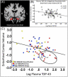
Plasma TDP-43 levels are associated with neuroimaging measures of brain structure in limbic regions
- PMID: 37266411
- PMCID: PMC10230689
- DOI: 10.1002/dad2.12437
Plasma TDP-43 levels are associated with neuroimaging measures of brain structure in limbic regions
Abstract
Introduction: We evaluated the relationship between plasma levels of transactive response DNA binding protein of 43 kDa (TDP-43) and neuroimaging (magnetic resonance imaging [MRI]) measures of brain structure in aging.
Methods: Plasma samples were collected from 72 non-demented older adults (age range 60-94 years) in the University of Kentucky Alzheimer's Disease Research Center cohort. Multivariate linear regression models were run with plasma TDP-43 level as the predictor variable and brain structure (volumetric or cortical thickness) measurements as the dependent variable. Covariates included age, sex, intracranial volume, and plasma markers of Alzheimer's disease neuropathological change (ADNC).
Results: Negative associations were observed between plasma TDP-43 level and both the volume of the entorhinal cortex, and cortical thickness in the cingulate/parahippocampal gyrus, after controlling for ADNC plasma markers.
Discussion: Plasma TDP-43 levels may be directly associated with structural MRI measures. Plasma TDP-43 assays may prove useful in clinical trial stratification.
Highlights: Plasma transactive response DNA binding protein of 43 kDa (TDP-43) levels were associated with entorhinal cortex volume.Biomarkers of TDP-43 and Alzheimer's disease neuropathologic change (ADNC) may help distinguish limbic-predominant age-related TDP-43 encephalopathy neuropathologic change (LATE-NC) from ADNC.A comprehensive biomarker kit could aid enrollment in LATE-NC clinical trials.
Keywords: aging; biomarker; cortical thickness; entorhinal cortex; limbic‐predominant age‐related TDP‐43 encephalopathy; neuroimaging; plasma transactive response DNA binding protein of 43 kDa.
© 2023 The Authors. Alzheimer's & Dementia: Diagnosis, Assessment & Disease Monitoring published by Wiley Periodicals, LLC on behalf of Alzheimer's Association.
Conflict of interest statement
The authors report no conflicts of interest. Author disclosures are available in the supporting information.
Figures


Similar articles
-
Cognitive symptoms progress with limbic-predominant age-related TDP-43 encephalopathy stage and co-occurrence with Alzheimer disease.J Neuropathol Exp Neurol. 2023 Dec 22;83(1):2-10. doi: 10.1093/jnen/nlad098. J Neuropathol Exp Neurol. 2023. PMID: 37966908 Free PMC article.
-
Neuropsychiatric symptoms in limbic-predominant age-related TDP-43 encephalopathy and Alzheimer's disease.Brain. 2020 Dec 1;143(12):3842-3849. doi: 10.1093/brain/awaa315. Brain. 2020. PMID: 33188391 Free PMC article.
-
Limbic Predominant Age-Related TDP-43 Encephalopathy (LATE): Clinical and Neuropathological Associations.J Neuropathol Exp Neurol. 2020 Mar 1;79(3):305-313. doi: 10.1093/jnen/nlz126. J Neuropathol Exp Neurol. 2020. PMID: 31845964 Free PMC article.
-
Structural magnetic resonance imaging for the early diagnosis of dementia due to Alzheimer's disease in people with mild cognitive impairment.Cochrane Database Syst Rev. 2020 Mar 2;3(3):CD009628. doi: 10.1002/14651858.CD009628.pub2. Cochrane Database Syst Rev. 2020. PMID: 32119112 Free PMC article.
-
The influence of Aβ-dependent and independent pathways on TDP-43 proteinopathy in Alzheimer's disease: a possible connection to LATE-NC.Neurobiol Aging. 2020 Nov;95:161-167. doi: 10.1016/j.neurobiolaging.2020.06.020. Epub 2020 Jul 2. Neurobiol Aging. 2020. PMID: 32814257 Review.
Cited by
-
Neuronal Vulnerability of the Entorhinal Cortex to Tau Pathology in Alzheimer's Disease.Br J Biomed Sci. 2024 Oct 7;81:13169. doi: 10.3389/bjbs.2024.13169. eCollection 2024. Br J Biomed Sci. 2024. PMID: 39435008 Free PMC article. Review.
-
Synergistic effects of plasma S100B and MRI measures of cerebrovascular disease on cognition in older adults.Geroscience. 2025 Jun;47(3):3131-3146. doi: 10.1007/s11357-024-01498-1. Epub 2025 Feb 5. Geroscience. 2025. PMID: 39907937 Free PMC article.
References
-
- Nelson PT, Brayne C, Flanagan ME, et al. Frequency of LATE neuropathologic change across the spectrum of Alzheimer's disease neuropathology: combined data from 13 community‐based or population‐based autopsy cohorts. Acta Neuropathol. 2022;144:27‐44. doi: 10.1007/s00401-022-02444-1 - DOI - PMC - PubMed
Grants and funding
LinkOut - more resources
Full Text Sources
