. 2023 Jun 2;18(6):e0282939.
doi: 10.1371/journal.pone.0282939. eCollection 2023.

Integral assays of hemostasis in hospitalized patients with COVID-19 on admission and during heparin thromboprophylaxis

Affiliations

Integral assays of hemostasis in hospitalized patients with COVID-19 on admission and during heparin thromboprophylaxis

Andrey Y Bulanov et al. PLoS One. .

Abstract

Background: Blood coagulation abnormalities play a major role in COVID-19 pathophysiology. However, the specific details of hypercoagulation and anticoagulation treatment require investigation. The aim of this study was to investigate the status of the coagulation system by means of integral and local clotting assays in COVID-19 patients on admission to the hospital and in hospitalized COVID-19 patients receiving heparin thromboprophylaxis.

Methods: Thrombodynamics (TD), thromboelastography (TEG), and standard clotting assays were performed in 153 COVID-19 patients observed in a hospital setting. All patients receiving treatment, except extracorporeal membrane oxygenation (ECMO) patients (n = 108), were administered therapeutic doses of low molecular weight heparin (LMWH) depending on body weight. The ECMO patients (n = 15) were administered unfractionated heparin (UFH).

Results: On admission, the patients (n = 30) had extreme hypercoagulation by all integral assays: TD showed hypercoagulation in ~75% of patients, while TEG showed hypercoagulation in ~50% of patients. The patients receiving treatment showed a significant heparin response based on TD; 77% of measurements were in the hypocoagulation range, 15% were normal, and 8% remained in hypercoagulation. TEG showed less of a response to heparin: 24% of measurements were in the hypocoagulation range, 59% were normal and 17% remained in hypercoagulation. While hypocoagulation is likely due to heparin treatment, remaining in significant hypercoagulation may indicate insufficient anticoagulation for some patients, which is in agreement with our clinical findings. There were 3 study patients with registered thrombosis episodes, and all were outside the target range for TD parameters typical for effective thromboprophylaxis (1 patient was in weak hypocoagulation, atypical for the LMWH dose used, and 2 patients remained in the hypercoagulation range despite therapeutic LMWH doses).

Conclusion: Patients with COVID-19 have severe hypercoagulation, which persists in some patients receiving anticoagulation treatment, while significant hypocoagulation is observed in others. The data suggest critical issues of hemostasis balance in these patients and indicate the potential importance of integral assays in its control.


Conflict of interest statement

The authors have declared that no competing interests exist.

Figures
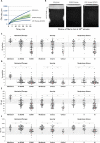
Fig 1
Fig 1. Thrombodynamics assay parameters in the patient groups.
(A) Examples of clot size as a function of time curves for patients on admission and during treatment and (B) images of clots for representative experiments. (C) Initial clot growth rate Vi, (D) stationary clot growth rate Vs, (E) clot size CS, and (F) clot density D in the patient groups: 1) on admission, with and without extracorporeal membrane oxygenation (ECMO) treatment; 2) depending on patient severity; and 3) depending on the severity of respiratory failure. The patterned area shows the normal reference ranges. The box plot indicates the following parameters: the median (the horizontal line inside the box), the 25th and 75th percentiles (the bottom and top of the box, respectively), and the 5th and 95th percentiles (the ends of the whiskers); * statistically significant difference (Mann‒Whitney U-criterion, p<0.01). NS represents no significant difference. Red circle symbol–bleeding episodes followed in patients receiving heparin therapy, red square symbol–bleeding episode followed in a patient with temporarily suspended heparin therapy, blue triangle symbol–thrombotic episodes followed in patients receiving heparin therapy.
Fig 2
Fig 2
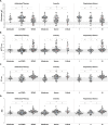
a. Thromboelastography (citrated native) parameters in the patient groups. (A) R, (B) K, (C) Angle, and (D) MA in the patient groups: 1) on admission, with and without extracorporeal membrane oxygenation (ECMO) treatment; 2) depending on patient severity; and 3) depending on the severity of respiratory failure. Note that TEG for patients on admission and for those without ECMO was performed with the citrated native version, while TEG for those with ECMO was performed with the native kaolin version. The patterned area shows the normal reference ranges. The box plot indicates the following parameters: the median (the horizontal line inside the box), the 25th and 75th percentiles (the bottom and top of the box, respectively), and the 5th and 95th percentiles (the ends of the whiskers); * statistically significant difference (Mann‒Whitney U-criterion, p<0.01). NS represents no significant difference. Red circle symbol–bleeding episodes followed in patients receiving heparin therapy, red square symbol–bleeding episode followed in a patient with temporarily suspended heparin therapy, blue triangle symbol–thrombotic episodes followed in patients receiving heparin therapy. b. Thromboelastography (citrated kaolin) parameters in the patient groups. (A) R, (B) K, (C) Angle, and (D) MA in the patient groups: 1) on admission, with and without extracorporeal membrane oxygenation (ECMO) treatment; 2) depending on patient severity; and 3) depending on the severity of respiratory failure. Note that TEG for patients on admission and for those without ECMO was performed with the citrated native version, while TEG for those with ECMO was performed with the native kaolin version. The patterned area shows the normal reference ranges. The box plot indicates the following parameters: the median (the horizontal line inside the box), the 25th and 75th percentiles (the bottom and top of the box, respectively), and the 5th and 95th percentiles (the ends of the whiskers); * statistically significant difference (Mann‒Whitney U-criterion, p<0.01). NS represents no significant difference. Red circle symbol–bleeding episodes followed in patients receiving heparin therapy, red square symbol–bleeding episode followed in a patient with temporarily suspended heparin therapy, blue triangle symbol–thrombotic episodes followed in patients receiving heparin therapy.
Fig 3
Fig 3. Clotting assays.
(A) Fibrinogen, (B) APTT, (C) PT, and (D) INR in the patient groups: 1) on admission, with and without extracorporeal membrane oxygenation (ECMO) treatment; 2) depending on patient severity; and 3) depending on the severity of respiratory failure. The patterned area shows the normal reference ranges. The box plot indicates the following parameters: the median (the horizontal line inside the box), the 25th and 75th percentiles (the bottom and top of the box, respectively), and the 5th and 95th percentiles (the ends of the whiskers); * statistically significant difference (Mann‒Whitney U-criterion, p<0.01). NS represents no significant difference. Red circle symbol–bleeding episodes followed in patients receiving heparin therapy, red square symbol–bleeding episode followed in a patient with temporarily suspended heparin therapy, blue triangle symbol–thrombotic episodes followed in patients receiving heparin therapy.
Fig 4
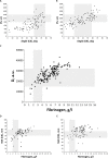
Fig 4. Correlation plots.
(A) TD CS vs. TEG Angle (citrated kaolin), (B) TD CS vs. TEG Angle (citrated native), (C) TD D vs. fibrinogen, (D) TEG MA (citrated kaolin) vs. fibrinogen, and (E) TEG MA (citrated native) vs. fibrinogen. The patterned area shows the normal reference ranges.

References

    1. Tang N, Li D, Wang X, Sun Z. Abnormal coagulation parameters are associated with poor prognosis in patients with novel coronavirus pneumonia. J Thromb Haemost. 2020;18: 844–847. doi: 10.1111/jth.14768 - DOI - PMC - PubMed
    1. Wu C, Chen X, Cai Y, Xia J, Zhou X, Xu S, et al.. Risk Factors Associated With Acute Respiratory Distress Syndrome and Death in Patients With Coronavirus Disease 2019 Pneumonia in Wuhan, China. JAMA Intern Med. 2020;180: 934–943. doi: 10.1001/jamainternmed.2020.0994 - DOI - PMC - PubMed
    1. Thomas MR, Scully M. Clinical features of thrombosis and bleeding in COVID-19. Blood. 2022;140: 184–195. doi: 10.1182/blood.2021012247 - DOI - PMC - PubMed
    1. Tan BK, Mainbourg S, Friggeri A, Bertoletti L, Douplat M, Dargaud Y, et al.. Arterial and venous thromboembolism in COVID-19: a study-level meta-analysis. Thorax. 2021;76: 970–979. doi: 10.1136/thoraxjnl-2020-215383 - DOI - PubMed
    1. Khismatullin RR, Ponomareva AA, Nagaswami C, Ivaeva RA, Montone KT, Weisel JW, et al.. Pathology of lung-specific thrombosis and inflammation in COVID-19. J Thromb Haemost. 2021;19: 3062–3072. doi: 10.1111/jth.15532 - DOI - PMC - PubMed

Publication types