Novel limbal dermoid surgery for visual acuity and cosmesis improvement: A 7-year retrospective review
- PMID: 37267334
- PMCID: PMC10237392
- DOI: 10.1371/journal.pone.0286250
Novel limbal dermoid surgery for visual acuity and cosmesis improvement: A 7-year retrospective review
Abstract
Background: To report a long-term outcome of the novel combined surgical method of complete excision, corneal tattooing, and a sutureless limbal conjunctival autograft for limbal dermoid.
Methods: All patients who were referred to our clinic for limbal dermoid, and underwent a combined surgery of complete excision, corneal tattooing, and a sutureless limbal conjunctival autograft were retrospectively reviewed. The surgery was performed by one surgeon, and all clinical information was obtained during a seven-year follow up period. In all patients, surgical outcomes of cosmesis, best corrected visual acuity (BCVA), spherical equivalent (SE), and corneal/ocular astigmatism were obtained and compared preoperatively and postoperatively.
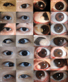
Results: During seven years, 24 patients (24 eyes) with limbal dermoid were finally enrolled. The mean age was 10.1±8.9 years old. The surgery resulted in an improved appearing ocular surface in all cases without any complications. There was no statistical difference in BCVA, corneal and ocular astigmatism between preoperatively and postoperatively (p = 0.231, 0.156 and 0.475, respectively). The mean SE was 0.12±3.19D preoperatively, and -0.21±3.02 D postoperatively with statistical significance (p = 0.037). Mean follow up period was 54.50 ± 15.62 months.
Conclusions: Based on the results of this study, our innovative surgical method which includes complete excision with corneal tattooing and limbal conjunctival autograft can be a simple and safe procedure that achieves long standing cosmesis with limbal dermoids.
Copyright: © 2023 Jeong et al. This is an open access article distributed under the terms of the Creative Commons Attribution License, which permits unrestricted use, distribution, and reproduction in any medium, provided the original author and source are credited.
Conflict of interest statement
The authors have declared that no competing interests exist.
Figures


References
-
- Mann I. Developmental Abnormalities of the Eye. 2n ed. Philadelphia, PA: British Medical Association; 1957; 74–91.
-
- Foster JA, Kherani F. Aesthetic Considerations in Pediatric Oculoplastic Surgery. Pediatric Oculoplastic Surgery. 2017; 279–290. doi: 10.1007/978-3-319-60814-3_18 - DOI
MeSH terms
LinkOut - more resources
Full Text Sources
Medical

