. 2023 Jun 2;9(22):eadg1082.
doi: 10.1126/sciadv.adg1082. Epub 2023 Jun 2.

Immune engineered extracellular vesicles to modulate T cell activation in the context of type 1 diabetes

Affiliations

Immune engineered extracellular vesicles to modulate T cell activation in the context of type 1 diabetes

Matthew W Becker et al. Sci Adv. .

Abstract

Extracellular vesicles (EVs) can affect immune responses through antigen presentation and costimulation or coinhibition. We generated designer EVs to modulate T cells in the context of type 1 diabetes, a T cell-mediated autoimmune disease, by engineering a lymphoblast cell line, K562, to express HLA-A*02 (HLA-A2) alongside costimulatory CD80 and/or coinhibitory programmed death ligand 1 (PD-L1). EVs presenting HLA-A2 and CD80 activated CD8+ T cells in a dose, antigen, and HLA-specific manner. Adding PD-L1 to these EVs produced an immunoregulatory response, reducing CD8+ T cell activation and cytotoxicity in vitro. EVs alone could not stimulate T cells without antigen-presenting cells. EVs lacking CD80 were ineffective at modulating CD8+ T cell activation, suggesting that both peptide-HLA complex and costimulation are required for EV-mediated immune modulation. These results provide mechanistic insight into the rational design of EVs as a cell-free approach to immunotherapy that can be tailored to promote inflammatory or tolerogenic immune responses.


Figures

Fig. 1.
Fig. 1.. EV isolation and characterization.
(A) Schematic overview of the workflow. Conditioned medium is processed through low-speed centrifugation and vacuum filtration to remove cells and cellular debris and then filtered through 300-kDa molecular weight cutoff (MWCO) UF devices to remove soluble proteins and concentrate the EV fraction. Concentrated medium is loaded onto a BE-SEC column using the ÄKTA Pure chromatography system. The first eluting fractions (EVs) are collected via an automated fraction collector and then subsequently concentrated using 100-kDa MWCO UF devices and used for further experiments. Contaminating proteins trapped by the BE-SEC column are eluted out during the column wash. (B) NTA showing representative particle concentration and size distribution of EVs derived from K562 A2/PPI cells. (C) Western blot analysis of EV isolates from K562 A2/PPI cells demonstrating the presence of small EV markers. n = 3. (D) Representative TEM image of K562 A2/PPI cell–derived EVs. Scale bar, 200 nm.
Fig. 2.
Fig. 2.. Validating an in vitro target:effector coculture model for EV engineering.
(A) Schematic representation of J-1E6 and K562 coculture. Yellow and gray circles represent endogenous peptides presented in HLA-A2 and exogenous PPI15–24 added into the coculture system, respectively. (B) IL-2 secretion from J-1E6 T cells showing activation from K562 A2/PPI cells, which is increased with exogenous peptide loading in HLA-A2. K562 cells lacking HLA-A2 fail to induce IL-2 secretion from J-1E6 T cells even in the presence of exogenous peptide. n = 3. Representative of four independent experiments. (C) Surface expression of the activation marker CD69 on J-1E6 T cells over a 24-hour coculture with K562 A2/PPI cells. n = 5. (D) Surface expression of PD-1 on J-1E6 T cells over a 24-hour coculture with K562 A2/PPI cells. n = 5. Statistical differences for (B) were determined by one-way analysis of variance (ANOVA) followed by Tukey’s multiple comparisons test. Statistical differences for (C) and (D) were determined by two-way ANOVA followed by Tukey’s multiple comparisons test. **P < 0.01, ***P < 0.001, ****P < 0.0001. ns, nonsignificant. (A) was created using BioRender.com. MFI, median fluorescent intensity.
Fig. 3.
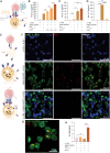
Fig. 3.. EVs need APCs to modulate T cell activation in vitro.
(A) Schematic representation of J-1E6 coculture with K562 A2/PPI cells and K-EVs. (B) IL-2 secretion from J-1E6 T cells showing increased activation with increasing amounts of K-EVs in cultures. Representative of five independent experiments. (C) IL-2 secretion from J-1E6 T cells after coculture with K562 A2/PPI cells and/or EVs, showing that EVs alone do not activate T cells. n = 3. (D) IL-2 secretion from J-1E6 cells after coculture with K562 A2/PPI cells or EVs, showing that EVs in the presence of exogenous PPI15–24 do not activate T cells. Representative of four independent experiments. (E) Schematic representations of the different conditions used in (F) to determine EV interactions in the coculture system. (F) Confocal microscopy images of DiR-labeled K-EVs incubated with either DiI-labeled J-1E6, DiO-labeled K562 A2/PPI, or both cell types for 24 hours. DiR signal was detected in cultures with K562 A2/PPI cells and J-1E6 with K562 A2/PPI cells but not J-1E6 cells alone, suggesting that K562 A2/PPI cells endocytose EVs. Representative of two independent experiments. (G) A high-resolution, three-dimensional micrograph of K562 A2/PPI cells incubated with DiR-labeled K-EVs. Yellow arrows indicate cell surface–bound EV clusters. (H) MFI quantification of confocal microscopy images, showing higher DiR EV signal in K562 A2/PPI cells and no significant difference between J-1E6 cells with or without K-EV coincubation. Each data point represents quantification from one image, with three images taken from each of two wells. Statistical differences for (B) through (D) and (H) were determined by one-way ANOVA followed by Tukey’s multiple comparisons test. *P < 0.05, **P < 0.01, ****P < 0.0001. ns, nonsignificant. (A) and (E) were created using BioRender.com. MFI, mean fluorescent intensity.
Fig. 4.
Fig. 4.. Driving PD-L1 expression in EV parent cells.
(A) Surface expression of PD-L1 on lentivirus-transduced cells shown via flow cytometry and further confirmed by Western blot analysis. (B) Schematic representation of J-1E6 coculture with either K562 A2/PPI or K562 PD-L1 cells, where PD-L1 engages with PD-1 to suppress IL-2 secretion. (C) IL-2 secretion from J-1E6 T cells showing decreased activation when cocultured with K562 PD-L1 cells compared to K562 A2/PPI. This suppression is still seen when exogenous PPI15–24 is added to cocultures. Representative of nine independent experiments. Statistical differences for (C) were determined by one-way ANOVA followed by Tukey’s multiple comparisons test. *P < 0.05, **P < 0.01. (B) was created using BioRender.com.
Fig. 5.
Fig. 5.. Validating PD-L1 expression in EVs.
(A) NTA showing representative particle concentration and size distribution of EVs derived from K562 PD-L1 cells. The inset shows the mode particle diameter for multiple K-EV and K–PD-L1–EV preparations, indicating that lentiviral transduction did not negatively affect EV generation. n = 13 and n = 12. (B) ELISA of PD-L1 on EVs from K562 cells compared to total EV protein content. n = 5. Data from two independent experiments. Statistical differences for (A) and (B) were determined by an unpaired, two-tailed t test. *P < 0.05. ns, nonsignificant.
Fig. 6.
Fig. 6.. K–PD-L1–EVs suppress T cell activation.
(A) Schematic representation of J-1E6 cocultures with K562 A2/PPI cells and KPD-L1EVs. (B) IL-2 secretion from J-1E6 cells showing decreased activation with K562 PD-L1 cells or increasing amounts of KPD-L1EVs in cultures, n = 3. Data shown are representative of three independent experiments. (C) Fold change in IL-2 secretion across all experiments with different EV treatments, showing that K-EVs significantly increase and KPD-L1EVs significantly decrease IL-2 secretion. T-EVs and TPD-L1EVs have no significant effect. Each dot represents a technical replicate from an independent experiment, and each color represents an independent experiment. Each technical replicate was normalized to its independent experimental control with just J-1E6 and K562 A2/PPI cells. Statistical differences for (B) were determined by one-way ANOVA followed by Tukey’s multiple comparisons test. Statistical differences for (C) were determined using a linear mixed effects model where fold change in IL-2 is predicted by the EV treatment as a linear fixed effect and the individual experiment as a random effect. *P < 0.05, **P < 0.01, ***P < 0.001, ****P < 0.0001. ns, nonsignificant. (A) was created using BioRender.com.
Fig. 7.
Fig. 7.. EV PD-L1 reduces T cell–mediated target cell killing in vitro.
(A) Schematic representation of T-IGRP cocultures with K562 A2/PPI cells and K–PD-L1–EVs. (B) Specific lysis of K562 cells by T-IGRP avatars, showing a 60% reduction in specific lysis of K562 A2/PPI cells when K–PD-L1–EVs are added to cocultures, n = 3. Representative of two independent experiments. (C) Specific lysis of K562 A2/PPI cells by T-IGRP avatars, showing a 60 to 70% increase in specific lysis when K-EVs are added to cocultures. Representative of two independent experiments. Statistical differences for (B) were determined by one-way ANOVA followed by Tukey’s multiple comparisons test. Statistical differences for (C) were determined by unpaired, two-tailed t test. *P < 0.05, ***P < 0.001, ****P < 0.0001. (A) was created using BioRender.com.

References

    1. van Niel G., D'Angelo G., Raposo G., Shedding light on the cell biology of extracellular vesicles. Nat. Rev. Mol. Cell Biol. 19, 213–228 (2018). - PubMed
    1. Thery C., Witwer K. W., Aikawa E., Alcaraz M. J., Anderson J. D., Andriantsitohaina R., Antoniou A., Arab T., Archer F., Atkin-Smith G. K., Ayre D. C., Bach J. M., Bachurski D., Baharvand H., Balaj L., Baldacchino S., Bauer N. N., Baxter A. A., Bebawy M., Beckham C., Zavec A. B., Benmoussa A., Berardi A. C., Bergese P., Bielska E., Blenkiron C., Bobis-Wozowicz S., Boilard E., Boireau W., Bongiovanni A., Borras F. E., Bosch S., Boulanger C. M., Breakefield X., Breglio A. M., Brennan M. A., Brigstock D. R., Brisson A., Broekman M. L., Bromberg J. F., Bryl-Gorecka P., Buch S., Buck A. H., Burger D., Busatto S., Buschmann D., Bussolati B., Buzas E. I., Byrd J. B., Camussi G., Carter D. R., Caruso S., Chamley L. W., Chang Y. T., Chen C., Chen S., Cheng L., Chin A. R., Clayton A., Clerici S. P., Cocks A., Cocucci E., Coffey R. J., Cordeiro-da-Silva A., Couch Y., Coumans F. A., Coyle B., Crescitelli R., Criado M. F., D'Souza-Schorey C., Das S., Chaudhuri A. D., de Candia P., De Santana E. F., De Wever O., Del Portillo H. A., Demaret T., Deville S., Devitt A., Dhondt B., Di Vizio D., Dieterich L. C., Dolo V., Rubio A. P. D., Dominici M., Dourado M. R., Driedonks T. A., Duarte F. V., Duncan H. M., Eichenberger R. M., Ekstrom K., El Andaloussi S., Elie-Caille C., Erdbrugger U., Falcon-Perez J. M., Fatima F., Fish J. E., Flores-Bellver M., Forsonits A., Frelet-Barrand A., Fricke F., Fuhrmann G., Gabrielsson S., Gamez-Valero A., Gardiner C., Gartner K., Gaudin R., Gho Y. S., Giebel B., Gilbert C., Gimona M., Giusti I., Goberdhan D. C., Gorgens A., Gorski S. M., Greening D. W., Gross J. C., Gualerzi A., Gupta G. N., Gustafson D., Handberg A., Haraszti R. A., Harrison P., Hegyesi H., Hendrix A., Hill A. F., Hochberg F. H., Hoffmann K. F., Holder B., Holthofer H., Hosseinkhani B., Hu G., Huang Y., Huber V., Hunt S., Ibrahim A. G., Ikezu T., Inal J. M., Isin M., Ivanova A., Jackson H. K., Jacobsen S., Jay S. M., Jayachandran M., Jenster G., Jiang L., Johnson S. M., Jones J. C., Jong A., Jovanovic-Talisman T., Jung S., Kalluri R., Kano S. I., Kaur S., Kawamura Y., Keller E. T., Khamari D., Khomyakova E., Khvorova A., Kierulf P., Kim K. P., Kislinger T., Klingeborn M., Klinke D. J. II, Kornek M., Kosanovic M. M., Kovacs A. F., Kramer-Albers E. M., Krasemann S., Krause M., Kurochkin I. V., Kusuma G. D., Kuypers S., Laitinen S., Langevin S. M., Languino L. R., Lannigan J., Lasser C., Laurent L. C., Lavieu G., Lazaro-Ibanez E., Le Lay S., Lee M. S., Lee Y. X. F., Lemos D. S., Lenassi M., Leszczynska A., Li I. T., Liao K., Libregts S. F., Ligeti E., Lim R., Lim S. K., Line A., Linnemannstons K., Llorente A., Lombard C. A., Lorenowicz M. J., Lorincz A. M., Lotvall J., Lovett J., Lowry M. C., Loyer X., Lu Q., Lukomska B., Lunavat T. R., Maas S. L., Malhi H., Marcilla A., Mariani J., Mariscal J., Martens-Uzunova E. S., Martin-Jaular L., Martinez M. C., Martins V. R., Mathieu M., Mathivanan S., Maugeri M., McGinnis L. K., McVey M. J., Meckes D. G. Jr., Meehan K. L., Mertens I., Minciacchi V. R., Moller A., Jorgensen M. M., Morales-Kastresana A., Morhayim J., Mullier F., Muraca M., Musante L., Mussack V., Muth D. C., Myburgh K. H., Najrana T., Nawaz M., Nazarenko I., Nejsum P., Neri C., Neri T., Nieuwland R., Nimrichter L., Nolan J. P., Nolte-'t Hoen E. N., Hooten N. N., O'Driscoll L., O'Grady T., O'Loghlen A., Ochiya T., Olivier M., Ortiz A., Ortiz L. A., Osteikoetxea X., Ostergaard O., Ostrowski M., Park J., Pegtel D. M., Peinado H., Perut F., Pfaffl M. W., Phinney D. G., Pieters B. C., Pink R. C., Pisetsky D. S., von Strandmann E. P., Polakovicova I., Poon I. K., Powell B. H., Prada I., Pulliam L., Quesenberry P., Radeghieri A., Raffai R. L., Raimondo S., Rak J., Ramirez M. I., Raposo G., Rayyan M. S., Regev-Rudzki N., Ricklefs F. L., Robbins P. D., Roberts D. D., Rodrigues S. C., Rohde E., Rome S., Rouschop K. M., Rughetti A., Russell A. E., Saa P., Sahoo S., Salas-Huenuleo E., Sanchez C., Saugstad J. A., Saul M. J., Schiffelers R. M., Schneider R., Schoyen T. H., Scott A., Shahaj E., Sharma S., Shatnyeva O., Shekari F., Shelke G. V., Shetty A. K., Shiba K., Siljander P. R., Silva A. M., Skowronek A., Snyder O. L. II, Soares R. P., Sodar B. W., Soekmadji C., Sotillo J., Stahl P. D., Stoorvogel W., Stott S. L., Strasser E. F., Swift S., Tahara H., Tewari M., Timms K., Tiwari S., Tixeira R., Tkach M., Toh W. S., Tomasini R., Torrecilhas A. C., Tosar J. P., Toxavidis V., Urbanelli L., Vader P., van Balkom B. W., van der Grein S. G., Van Deun J., van Herwijnen M. J., Van Keuren-Jensen K., van Niel G., van Royen M. E., van Wijnen A. J., Vasconcelos M. H., Vechetti I. J. Jr., Veit T. D., Vella L. J., Velot E., Verweij F. J., Vestad B., Vinas J. L., Visnovitz T., Vukman K. V., Wahlgren J., Watson D. C., Wauben M. H., Weaver A., Webber J. P., Weber V., Wehman A. M., Weiss D. J., Welsh J. A., Wendt S., Wheelock A. M., Wiener Z., Witte L., Wolfram J., Xagorari A., Xander P., Xu J., Yan X., Yanez-Mo M., Yin H., Yuana Y., Zappulli V., Zarubova J., Zekas V., Zhang J. Y., Zhao Z., Zheng L., Zheutlin A. R., Zickler A. M., Zimmermann P., Zivkovic A. M., Zocco D., Zuba-Surma E. K., Minimal information for studies of extracellular vesicles 2018 (MISEV2018): A position statement of the International Society for Extracellular Vesicles and update of the MISEV2014 guidelines. J. Extracell Vesicles 7, 1535750 (2018). - PMC - PubMed
    1. Witwer K. W., Thery C., Extracellular vesicles or exosomes? On primacy, precision, and popularity influencing a choice of nomenclature. J. Extracell Vesicles 8, 1648167 (2019). - PMC - PubMed
    1. Kalluri R., LeBleu V. S., The biology, function, and biomedical applications of exosomes. Science 367, eaau6977 (2020). - PMC - PubMed
    1. Harmati M., Bukva M., Boroczky T., Buzas K., Gyukity-Sebestyen E., The role of the metabolite cargo of extracellular vesicles in tumor progression. Cancer Metastasis Rev. 40, 1203–1221 (2021). - PMC - PubMed

MeSH terms