Review
. 2023 Sep:126:103864.
doi: 10.1016/j.mcn.2023.103864. Epub 2023 Jun 1.

Consequences of oxygen deprivation on myelination and sex-dependent alterations

Affiliations
Review

Consequences of oxygen deprivation on myelination and sex-dependent alterations

Rafael Bandeira Fabres et al. Mol Cell Neurosci. 2023 Sep.

Abstract

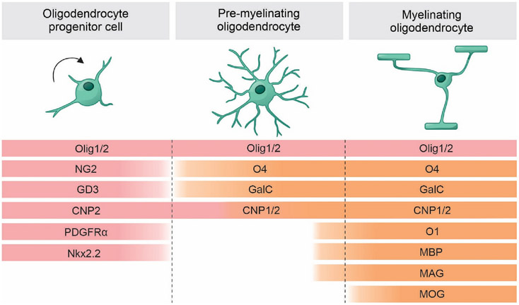
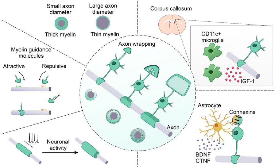
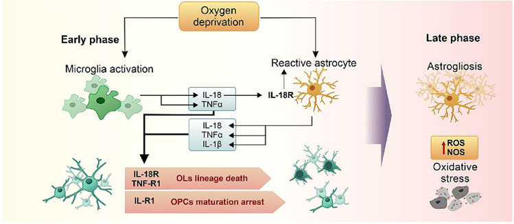
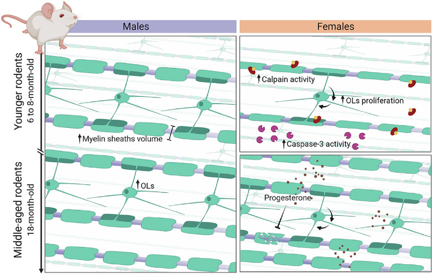
Oxygen deprivation is one of the main causes of morbidity and mortality in newborns, occurring with a higher prevalence in preterm infants, reaching 20 % to 50 % mortality in newborns in the perinatal period. When they survive, 25 % exhibit neuropsychological pathologies, such as learning difficulties, epilepsy, and cerebral palsy. White matter injury is one of the main features found in oxygen deprivation injury, which can lead to long-term functional impairments, including cognitive delay and motor deficits. The myelin sheath accounts for much of the white matter in the brain by surrounding axons and enabling the efficient conduction of action potentials. Mature oligodendrocytes, which synthesize and maintain myelination, also comprise a significant proportion of the brain's white matter. In recent years, oligodendrocytes and the myelination process have become potential therapeutic targets to minimize the effects of oxygen deprivation on the central nervous system. Moreover, evidence indicate that neuroinflammation and apoptotic pathways activated during oxygen deprivation may be influenced by sexual dimorphism. To summarize the most recent research about the impact of sexual dimorphism on the neuroinflammatory state and white matter injury after oxygen deprivation, this review presents an overview of the oligodendrocyte lineage development and myelination, the impact of oxygen deprivation and neuroinflammation on oligodendrocytes in neurodevelopmental disorders, and recent reports about sexual dimorphism regarding the neuroinflammation and white matter injury after neonatal oxygen deprivation.

Keywords: Myelination; Neuroinflammation; Oligodendrocytes; Oxygen deprivation; Sexual dimorphism; White matter injury.


Conflict of interest statement

Declaration of competing interest The authors declare that they have no known competing financial interests or personal relationships that could have appeared to influence the work reported in this paper.

Figures

Fig. 1.
Fig. 1.
Markers of oligodendrocyte developmental stages. Oligodendrocyte progenitor cells mark the initial stages of differentiation. In this stage, cells can be identified by the expression of NG2, PDGFRα, NKx2.2, CNP2 and GD3. Pre-myelinating period is characterized by the initial expression of mature markers, such as MBP, MAG, CNP1, O4, O1 and GalC. Myelinating period is characterized by the expression of MOG. Olig 1 and 2 are expressed throughout the lineage and support the maturation process. NG2: neuron-glial antigen 2; PDGFαR: platelet-derived growth factor receptor A; Nkx2.2: NK2 homeobox 2; CNP2: CNPase 2; CNP1: CNPase 1; Olig 1: oligodendrocyte transcription factor 1; Olig 2: oligodendrocyte transcription factor 2; GD3: GD3 ganglioside; MBP: myelin basic protein; MAG: myelin associated glycoprotein; MOG: myelin-oligodendrocyte glycoprotein; GalC: galactocerebroside.
Fig. 2.
Fig. 2.
CNS model of myelination and guidance factors of the myelination process. In the center, the current proposed model of myelination to the CNS is illustrated by an unmyelinated axon in parallel to an oligodendrocyte in three sequential steps of myelination and the respective cross-section illustration. In the first step, the triangle shape inner tongue is projected toward the axon, followed by the repeated encircle of the axon, lateral extension, and compaction of the myelin layers. Factors that guide the oligodendrocytes' decision to myelinate or not an axon are illustrated around the main image. Such factors include axon diameter, attractive or repulsive molecular cues, and neuronal activity. Moreover, the contribution of other glial cells is highlighted: astrocytes secrete BDNF and CTNF couple with oligodendrocytes by connexins; microglia CD11c + support the myelination process by the secretion of IGF-1, especially in the corpus callosum. IGF-1: insulin-like growth factor 1; BDNF: Brain-Derived Neurotrophic Factor; CTNF: ciliary neurotrophic factor.
Fig. 3.
Fig. 3.
Neuroinflammatory phases after neonatal oxygen deprivation. The schematic illustration of the early neuroinflammatory phase evidence the activation of the microglia and astrocytes especially by the interaction of the IL-18 secreted by the microglia with the IL-18R expressed by the astrocytes. Next, the interaction of IL-18, TNFα, and IL-1β released by microglia and astrocytes with their respective receptors expressed mainly in pre-myelinating OLs promotes death of the lineage or OPC maturation arrest, culminating in myelination deficits. In the latter phase, astrogliosis promotes the elevation of ROS and NOS levels; as immature OLs are susceptible to oxidative stress, later impairment of oligodendrocyte lineage continues.
Fig. 4.
Fig. 4.
Myelin differences among males and females throughout development. Males' myelination, in the left, is constant from younger through adulthood rodents, if the myelin sheath, the myelinated fibers volume, and the number of OLs are considered. In the right, if compared to younger males, the schematic illustration shows the higher calpain and caspase-3 activity, and faster turnover of OLs in females of the same age, which is reflected in the reduced myelin sheath and myelinate fibers volume, and number of OLs. In adulthood, females and males have comparable myelination, despite faster turnover in females. In this age, progesterone administration in females is a neuroprotector that restore demyelination lesions.

References

    1. Aberg ND, Johansson UE, Aberg MA, Hellstrom NA, Lind J, Bull C, Isgaard J, Anderson MF, Oscarsson J, Eriksson PS, 2007. Peripheral infusion of insulin-like growth factor-I increases the number of newborn oligodendrocytes in the cerebral cortex of adult hypophysectomized rats. Endocrinology 148, 3765–3772. - PubMed
    1. Arteni NS, Pereira LO, Rodrigues AL, Lavinsky D, Achaval ME, Netto CA, 2010. Lateralized and sex-dependent behavioral and morphological effects of unilateral neonatal cerebral hypoxia-ischemia in the rat. Behav. Brain Res 210, 92–98. - PubMed
    1. Back SA, 2006. Perinatal white matter injury: the changing spectrum of pathology and emerging insights into pathogenetic mechanisms. Ment. Retard. Dev. Disabil. Res. Rev 12, 129–140. - PubMed
    1. Back SA, Miller SP, 2014. Brain injury in premature neonates: a primary cerebral dysmaturation disorder? Ann. Neurol 75, 469–486. - PMC - PubMed
    1. Back SA, Luo NL, Borenstein NS, Levine JM, Volpe JJ, Kinney HC, 2001. Late oligodendrocyte progenitors coincide with the developmental window of vulnerability for human perinatal white matter injury. J. Neurosci 21, 1302–1312. - PMC - PubMed

Publication types

LinkOut - more resources