

Optimized SPECT Imaging of 224Ra α-Particle Therapy by 212Pb Photon Emissions
- PMID: 37268424
- PMCID: PMC10315694
- DOI: 10.2967/jnumed.122.264455
Optimized SPECT Imaging of 224Ra α-Particle Therapy by 212Pb Photon Emissions
Abstract
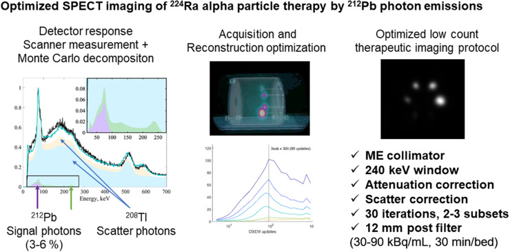
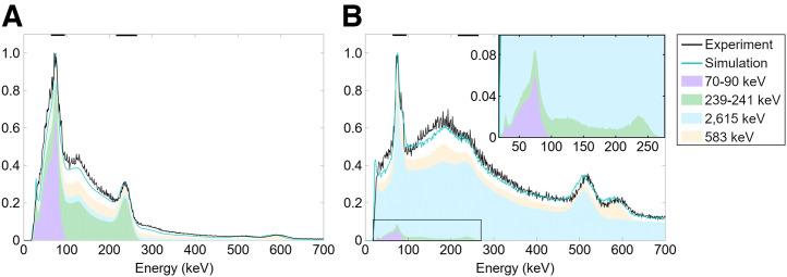
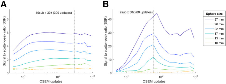
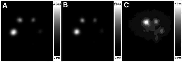
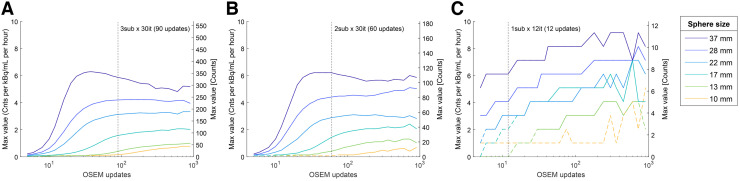
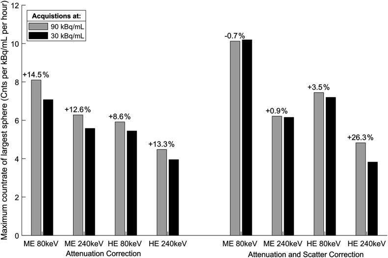
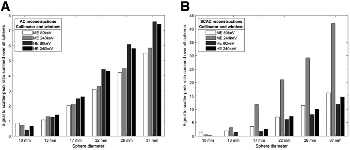
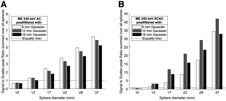
In preparation for an α-particle therapy trial using 1-7 MBq of 224Ra, the feasibility of tomographic SPECT/CT imaging was of interest. The nuclide decays in 6 steps to stable 208Pb, with 212Pb as the principle photon-emitting nuclide. 212Bi and 208Tl emit high-energy photons up to 2,615 keV. A phantom study was conducted to determine the optimal acquisition and reconstruction protocol. Methods: The spheres of a body phantom were filled with a 224Ra-RaCl2 solution, and the background compartment was filled with water. Images were acquired on a SPECT/CT system. In addition, 30-min scans were acquired for 80- and 240-keV emissions, using triple-energy windows, with both medium-energy and high-energy collimators. Images were acquired at 90-95 and 29-30 kBq/mL, plus an explorative 3-min acquisition at 20 kBq/mL (using only the optimal protocol). Reconstructions were performed with attenuation correction only, attenuation plus scatter correction, 3 levels of postfiltering, and 24 levels of iterative updates. Acquisitions and reconstructions were compared using the maximum value and signal-to-scatter peak ratio for each sphere. Monte Carlo simulations were performed to examine the contributions of key emissions. Results: Secondary photons of the 2,615-keV 208Tl emission produced in the collimators make up most of the acquired energy spectrum, as revealed by Monte Carlo simulations, with only a small fraction (3%-6%) of photons in each window providing useful information for imaging. Still, decent image quality is possible at 30 kBq/mL, and nuclide concentrations are imageable down to approximately 2-5 kBq/mL. The overall best results were obtained with the 240-keV window, medium-energy collimator, attenuation and scatter correction, 30 iterations and 2 subsets, and a 12-mm gaussian postprocessing filter. However, all combinations of the applied collimators and energy windows were capable of producing adequate results, even though some failed to reconstruct the 2 smallest spheres. Conclusion: SPECT/CT imaging of 224Ra in equilibrium with daughters is possible, with sufficient image quality to provide clinical utility for the current trial of intraperitoneally administrated activity. A systematic scheme for optimization was designed to select acquisition and reconstruction settings.
Keywords: Pb212; Ra224; SPECT; optimization; α-particle therapy.
© 2023 by the Society of Nuclear Medicine and Molecular Imaging.
Figures
Similar articles
-
Monte Carlo simulation study to explore optimum conditions for Astatine-211 SPECT.Radiol Phys Technol. 2023 Mar;16(1):102-108. doi: 10.1007/s12194-023-00702-9. Epub 2023 Jan 31. Radiol Phys Technol. 2023. PMID: 36719548
-
Evaluation the effect of different collimators and energy window on Y-90 bremsstrahlung SPECT imaging by SIMIND Monte Carlo program.Nucl Med Rev Cent East Eur. 2019;22(2):45-55. doi: 10.5603/NMR.a2019.0016. Nucl Med Rev Cent East Eur. 2019. PMID: 31482556
-
Feasibility and limitations of quantitative SPECT for 223Ra.Phys Med Biol. 2020 Apr 20;65(8):085012. doi: 10.1088/1361-6560/ab7971. Phys Med Biol. 2020. PMID: 32092708
-
Fast Monte Carlo based joint iterative reconstruction for simultaneous 99mTc/ 123I SPECT imaging.Med Phys. 2007 Aug;34(8):3263-72. doi: 10.1118/1.2756601. Med Phys. 2007. PMID: 17879789
-
Targeted Alpha Therapy: All We Need to Know about 225Ac's Physical Characteristics and Production as a Potential Theranostic Radionuclide.Pharmaceuticals (Basel). 2023 Dec 2;16(12):1679. doi: 10.3390/ph16121679. Pharmaceuticals (Basel). 2023. PMID: 38139806 Free PMC article. Review.
Cited by
-
Comparison of ZnS(Ag) Scintillator and Proportional Counter Tube for Alpha Detection in Thin-Layer Chromatography.Pharmaceuticals (Basel). 2024 Dec 28;18(1):26. doi: 10.3390/ph18010026. Pharmaceuticals (Basel). 2024. PMID: 39861089 Free PMC article.
-
Imaging of 212Pb in mice with a clinical SPECT/CT.EJNMMI Phys. 2023 Aug 21;10(1):47. doi: 10.1186/s40658-023-00571-6. EJNMMI Phys. 2023. PMID: 37603123 Free PMC article.
-
First-in-Human Phase 0 Study of AB001, a Prostate-Specific Membrane Antigen-Targeted 212Pb Radioligand, in Patients with Metastatic Castration-Resistant Prostate Cancer.J Nucl Med. 2025 May 1;66(5):732-738. doi: 10.2967/jnumed.124.269299. J Nucl Med. 2025. PMID: 40081958 Free PMC article.
-
Different 212Pb Generators and Its Radiation Safety Concerning 220Rn (Thoron) Emanation.Toxics. 2025 May 30;13(6):462. doi: 10.3390/toxics13060462. Toxics. 2025. PMID: 40559935 Free PMC article.
-
Theranostic Imaging Surrogates for Targeted Alpha Therapy: Progress in Production, Purification, and Applications.Pharmaceuticals (Basel). 2023 Nov 17;16(11):1622. doi: 10.3390/ph16111622. Pharmaceuticals (Basel). 2023. PMID: 38004486 Free PMC article. Review.
References
-
- Pacilio M, Ventroni G, De Vincentis G, et al. . Dosimetry of bone metastases in targeted radionuclide therapy with alpha-emitting (223)Ra-dichloride. Eur J Nucl Med Mol Imaging. 2016;43:21–33. - PubMed
-
- Hindorf C, Chittenden S, Aksnes AK, Parker C, Flux GD. Quantitative imaging of 223Ra-chloride (Alpharadin) for targeted alpha-emitting radionuclide therapy of bone metastases. Nucl Med Commun. 2012;33:726–732. - PubMed
-
- Sgouros G, Frey E, Du Y, Hobbs R, Bolch W. Imaging and dosimetry for alpha-particle emitter radiopharmaceutical therapy: improving radiopharmaceutical therapy by looking into the black box. Eur J Nucl Med Mol Imaging. 2021;49:18–29. - PubMed
-
- Cordier D, Forrer F, Bruchertseifer F, et al. . Targeted alpha-radionuclide therapy of functionally critically located gliomas with 213Bi-DOTA-[Thi8,Met(O2)11]-substance P: a pilot trial. Eur J Nucl Med Mol Imaging. 2010;37:1335–1344. - PubMed
MeSH terms
Substances
LinkOut - more resources
Full Text Sources
Other Literature Sources