. 2023 Jul 18;101(3):e289-e299.
doi: 10.1212/WNL.0000000000207397. Epub 2023 Jun 2.

Clinicoradiologic and Neuropathologic Evaluation of Corticobasal Syndrome

Affiliations

Clinicoradiologic and Neuropathologic Evaluation of Corticobasal Syndrome

Dror Shir et al. Neurology. .

Abstract

Background and objectives: Corticobasal syndrome (CBS) is a clinical phenotype characterized by asymmetric parkinsonism, rigidity, myoclonus, and apraxia. Originally believed secondary to corticobasal degeneration (CBD), mounting clinicopathologic studies have revealed heterogenous neuropathologies. The objectives of this study were to determine the pathologic heterogeneity of CBS, the clinicoradiologic findings associated with different underlying pathologies causing CBS, and the positive predictive value (PPV) of current diagnostic criteria for CBD among patients with a CBS.

Methods: Clinical data, brain MRI, and neuropathologic data of patients followed at Mayo Clinic and diagnosed with CBS antemortem were reviewed according to neuropathology category at autopsy.

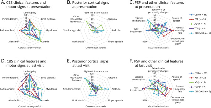
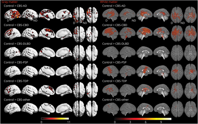
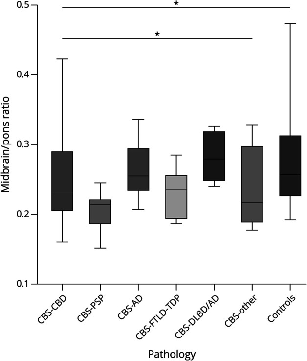
Results: The cohort consisted of 113 patients with CBS, 61 (54%) female patients. Mean ± SD disease duration was 7 ± 3.7 years; mean ± SD age at death was 70.5 ± 9.1 years. The primary neuropathologic diagnoses were 43 (38%) CBD, 27 (24%) progressive supranuclear palsy (PSP), 17 (15%) Alzheimer disease (AD), 10 (9%) frontotemporal lobar degeneration (FTLD) with TAR DNA-binding protein 43 (TDP) inclusions, 7 (6%) diffuse Lewy body disease (DLBD)/AD, and 9 (8%) with other diagnoses. Patients with CBS-AD or CBS-DLBD/AD were youngest at death (median [interquartile range]: 64 [13], 64 [11] years) while CBS-PSP were oldest (77 [12.5] years, p = 0.024). Patients with CBS-DLBD/AD had the longest disease duration (9 [6] years), while CBS-other had the shortest (3 [4.25] years, p = 0.04). Posterior cortical signs and myoclonus were more characteristic of patients with CBS-AD and patients with CBS-DLBD/AD. Patients with CBS-DLBD/AD displayed more features of Lewy body dementia. Voxel-based morphometry revealed widespread cortical gray matter loss characteristic of CBS-AD, while CBS-CBD and CBS-PSP predominantly involved premotor regions with greater amount of white matter loss. Patients with CBS-DLBD/AD showed atrophy in a focal parieto-occipital region, and patients with CBS-FTLD-TDP had predominant prefrontal cortical loss. Patients with CBS-PSP had the lowest midbrain/pons ratio (p = 0.012). Of 67 cases meeting clinical criteria for possible CBD at presentation, 27 were pathology-proven CBD, yielding a PPV of 40%.

Discussion: A variety of neurodegenerative disorders can be identified in patients with CBS, but clinical and regional imaging differences aid in predicting underlying neuropathology. PPV analysis of the current CBD diagnostic criteria revealed suboptimal performance. Biomarkers adequately sensitive and specific for CBD are needed.


Conflict of interest statement

D.S. Knopman serves on a Data Safety Monitoring Board for the DIAN study. He served on a Data Safety monitoring Board for a tau therapeutic for Biogen but receives no personal compensation. He is a site investigator in the Biogen aducanumab trials. He is an investigator in a clinical trial sponsored by Lilly Pharmaceuticals and the University of Southern California. He serves as a consultant for Samus Therapeutics, Roche, and Alzeca Biosciences but receives no personal compensation. R.C. Petersen serves as a consultant for Biogen, Inc., Roche, Inc., Merck, Inc., Genentech Inc. (DSMB), Nestle, Inc., and Eisai, Inc.; receives publishing royalties from Mild Cognitive Impairment (Oxford University Press, 2003) and UpToDate. B.F. Boeve, D.W. Dickson, K.A. Josephs, and J.L. Whitwell received research funding from the NIH and declare no competing financial interests. M.E. Murray is a consultant for AVID Radiopharmaceuticals. She receives support from the NIH/NIA and State of Florida. N.R. Graff-Radford receives royalties from UpToDate; has participated in multicenter therapy studies sponsored by Biogen, TauRx, AbbVie, Novartis, and Lilly; and he receives research support from NIH. J. Graff-Radford serves on the editorial board for Neurology® and receives research support from the NIH. The other authors declare no financial or other conflict of interest. Go to Neurology.org/N for full disclosures.

Figures

Figure 1
Figure 1. Radar Charts of Clinical Features by Neuropathology Diagnosis
(A–C) At presentation. (D–F) At last visit. Axes present percentage. Panel A: At presentation, myoclonus and apraxia were more frequent in CBS-AD (green, p = 0.003) and CBS-DLBD/AD (light blue, p = 0.017); alien limb phenomenon was more frequent in CBS-CBD (dark blue), CBS-PSP (red), and CBS-AD (green, p = 0.036). Panel B: At presentation, posterior cortical signs were more frequent in CBS-AD (green) and CBS-DLBD/AD (light blue, including agraphia [p = 0.002], acalculia [p = 0.003], simultanagnosia [p = 0.005], and other visuospatial features [p < 0.001]). Panel C: Visual hallucinations were more frequent in CBS-DLBD/AD (p = 0.003). Panel D: At last visit, myoclonus remained more frequent in CBS-AD (green) and CBS-DLBD/AD (blue, p = 0.006). Panel E: At last visit, the above mentioned posterior cortical signs remained more frequent among CBS-AD (green) and CBS-DLBD/AD (light blue), in addition to optic ataxia (p = 0.004). Panel F: At last visit, core features of LBD were more frequent in patients with CBS-DLBD/AD (including visual hallucinations [p = 0.01], RBD [p = 0.032]); episodic memory loss was more frequent among CBS-AD (green) and CBS-DLBD/AD (p < 0.001); typical PSP features were more frequent in CBS-PSP (red), and apraxia of speech was more frequent in CBS-CBD (dark blue), but these trends did not reach statistical significance. For Panels B and E: Other visuospatial features at presentation include visual field defect, space/object perception deficit, alexia, dressing apraxia, and prosopagnosia. For Panels A–C: CBD (n = 38), PSP (n = 26), AD (n = 17), FTLD-TDP43 (n = 8), DLBD/AD (n = 7). For Panels D–F: CBD (n = 26), PSP (n = 22), AD (n = 15), FTLD-TDP43 (n = 7), DLBD/AD (n = 5). AD = Alzheimer disease; CBD = corticobasal degeneration; CBS = corticobasal syndrome; DLBD = diffuse Lewy body dementia; LBD = Lewy body dementia; PSP = progressive supranuclear palsy; RBD = REM sleep behavior disorder; TDP = TAR DNA-binding protein 43.
Figure 2
Figure 2. Regions of Gray Matter Loss in Patients With CBS According to Pathology Category
All results are FWE-corrected for multiple comparisons, p < 0.05, extent threshold = 20. Legends show t scores with brighter colors representing higher score. N = 103 (24 CBD, 13 AD, 11 PSP, 7 TDP43, 4 DLBD/AD, 4 other, 40 controls). AD = Alzheimer disease; CBD = corticobasal degeneration; CBS = corticobasal syndrome; D = dominant; DLBD = diffuse Lewy body dementia; FWE = family-wise error; ND = nondominant; PSP = progressive supranuclear palsy; TDP = TAR DNA-binding protein 43.
Figure 3
Figure 3. FDG-PET Across CBS Pathology Categories
Panels A–E show Z scores, relative to a normative database, of pons intensity normalized FDG-PET scans for each individual displayed on stereotactic surface projections using Cortex ID (GE Healthcare, Waukesha, WI). Red color indicates greater hypometabolism. AD = Alzheimer disease; CBD = corticobasal degeneration; CBS = corticobasal syndrome; DLBD = diffuse Lewy body dementia; FDG = 18F fluorodeoxyglucose; PSP = progressive supranuclear palsy; TDP = TAR DNA-binding protein 43.
Figure 4
Figure 4. Boxplot Showing Midbrain/Pons Ratio Differences Across Pathology Category
The boxes indicate the median and interquartile range of the distributions while the horizontal lines extending from the boxes stop at the most extreme data points. p Values were calculated using the Kruskal-Wallis test (p = 0.012 across all groups, p = 0.028 without controls). N = 103 (24 CBD, 13 AD, 11 PSP, 7 FTLD-TDP, 4 DLBD/AD, 4 other, 40 controls). AD = Alzheimer disease; CBD = corticobasal degeneration; CBS = corticobasal syndrome; DLBD = diffuse Lewy body dementia; FTLD = frontotemporal lobar degeneration; PSP = progressive supranuclear palsy; TDP = TAR DNA-binding protein 43.

References

    1. Svenningsson P. Corticobasal degeneration: advances in clinicopathology and biomarkers. Curr Opin Neurol. 2019;32(4):597-603. doi:10.1097/wco.0000000000000707 - DOI - PubMed
    1. Lee SE, Rabinovici GD, Mayo MC, et al. . Clinicopathological correlations in corticobasal degeneration. Ann Neurol. 2011;70(2):327-340. doi:10.1002/ana.22424 - DOI - PMC - PubMed
    1. Whitwell JL, Jack CR, Boeve BF, et al. . Imaging correlates of pathology in corticobasal syndrome. Neurology. 2010;75(21):1879-1887. doi:10.1212/wnl.0b013e3181feb2e8 - DOI - PMC - PubMed
    1. Ling H, O'Sullivan SS, Holton JL, et al. . Does corticobasal degeneration exist? A clinicopathological re-evaluation. Brain. 2010;133(7):2045-2057. doi:10.1093/brain/awq123 - DOI - PubMed
    1. Boxer AL, Gold M, Feldman H, et al. . New directions in clinical trials for frontotemporal lobar degeneration: methods and outcome measures. Alzheimers Dement. 2020;16(1):131-143. doi:10.1016/j.jalz.2019.06.4956 - DOI - PMC - PubMed

Publication types