. 2023 Jun 3;23(1):501.
doi: 10.1186/s12885-023-10983-3.

High HSPB1 expression predicts poor clinical outcomes and correlates with breast cancer metastasis

Affiliations

High HSPB1 expression predicts poor clinical outcomes and correlates with breast cancer metastasis

Qin Huo et al. BMC Cancer. .

Abstract

Background: Heat shock protein beta-1 (HSPB1) is a crucial biomarker for pathological processes in various cancers. However, the clinical value and function of HSPB1 in breast cancer has not been extensively explored. Therefore, we adopted a systematic and comprehensive approach to investigate the correlation between HSPB1 expression and clinicopathological features of breast cancer, as well as determine its prognostic value. We also examined the effects of HSPB1 on cell proliferation, invasion, apoptosis, and metastasis.

Methods: We investigated the expression of HSPB1 in patients with breast cancer using The Cancer Genome Atlas and immunohistochemistry. Chi-squared test and Wilcoxon signed-rank test were used to examine the relationship between HSPB1 expression and clinicopathological characteristics.

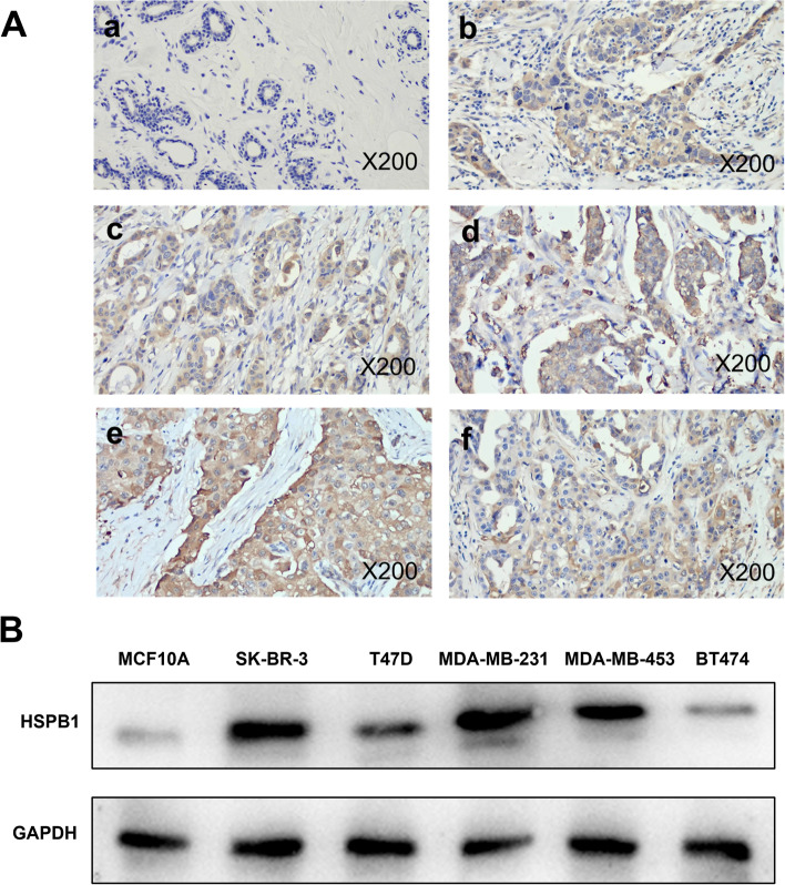
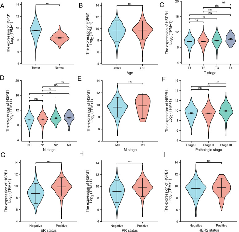
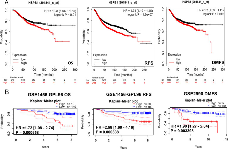
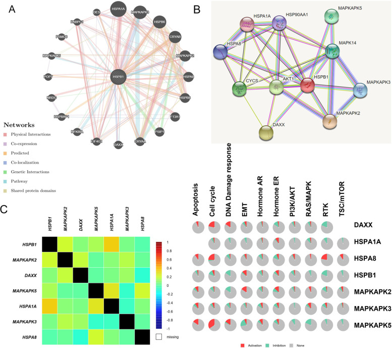
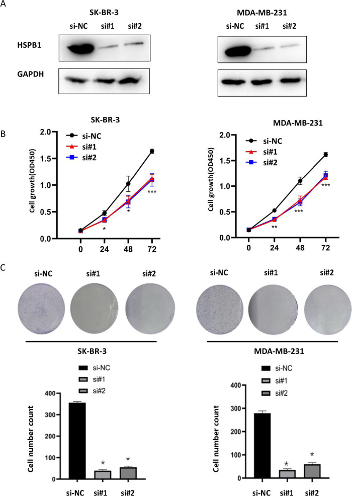
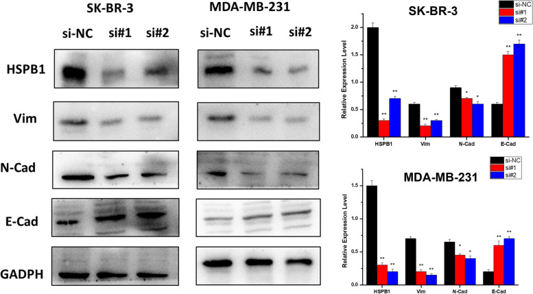
Results: We observed that HSPB1 expression was significantly correlated with the stage N, pathologic stages, as well as estrogen and progesterone receptors. Furthermore, high HSPB1 expression resulted in a poor prognosis for overall survival, relapse-free survival, and distant metastasis-free survival. Multivariable analysis showed that patients with poor survival outcomes had higher tumor, node, metastasis, and pathologic stages. Pathway analysis of HSPB1 and the altered neighboring genes suggested that HSPB1 is involved in the epithelial-to-mesenchymal transition. Functional analysis revealed showed that transient knockdown of HSPB1 inhibited the cell migration/invasion ability and promoted apoptosis.

Conclusions: HSPB1 may be involved in breast cancer metastasis. Collectively, our study demonstrated that HSPB1 has prognostic value for clinical outcomes and may serve as a therapeutic biomarker for breast cancer.

Keywords: Breast cancer; Cell proliferation; HSPB1; Metastasis; Prognostic biomarker.


Conflict of interest statement

The authors have no competing interests.

Figures

Fig. 1
Fig. 1
HSPB1 expression in breast cancer tissues and cells. A Immunohistochemical analysis of HSPB1 expression in breast tumor tissues (b-f) and normal tissues (a) (200 × magnification,). B Western blots showing the expression of HSPB1 protein in six breast cancer cell lines. The blots were cut prior to hybridisation with antibodies during blotting
Fig. 2
Fig. 2
Association between the HSPB1 expression levels and clinical characteristics in patients with breast cancer. The relationship between HSPB1 expression and clinical characteristics, including the patient A status (tumor or normal), B age (≤ 60 and > 60), C T stage (T1, T2, T3, or T4), D N stage (N0, N1, N2, or N3), E M stage (M0 or M1), F pathologic stage (stage I, stage II, or stage III), G estrogen receptor (ER; negative or positive), H progesterone receptor (PR; negative or positive). I human epidermal growth factor receptor 2 (HER2; negative or positive)
Fig. 3
Fig. 3
An analysis of the prognostic value of HSPB1. A Survival curves for overall survival (OS), relapse Free Survival (RFS), and distant metastasis free survival (DMFS) using the Kaplan –Meier plotter. B Survival curves for OS, RFS, and DMFS using the PrognoScan database
Fig. 4
Fig. 4
Identification of HSPB1-interacting genes and proteins and pathway analysis. The gene–gene interaction network and protein–protein interaction network of HSPB1 were constructed using GeneMANIA A and STRING B. C The correlation between HSPB1 and the altered neighboring genes using bc-GenExMiner v 4.8. D The GSCALite protocol was used to analyze the pathway activity (activation and inhibition)
Fig. 5
Fig. 5
Transient knockdown of HSPB1 affects breast cancer cell proliferation. A Verification of HSPB1 expression in SK-BR-3 and MDA-MB-231 cell lines via western blot. Growth curves B and Colony-forming efficiency C in SK-BR-3 and MDA-MB-231 cells before and after HSPB1 transient knockdown. The quantification of each analysis is shown in the following figure. All assays were performed in triplicate. The data are presented as means ± SEM. (*p < 0.05; **p < 0.01; *** p < 0.001). The blots were cut prior to hybridisation with antibodies during blotting
Fig. 6
Fig. 6
Transient knockdown of HSPB1 affects breast cancer cell migration, invasion, and apoptosis. A The effect of transient knockdown of HSPB1 expression on the cell migration was determined by using the wound healing assay. The quantification of each analysis is shown in the right figure. The experiments were carried out in triplicate (**p < 0.01). The scratch area was calculated using Image J software. Cell scratch area (0 h) minus cell scratch area (72 h) to get the cell migration area, the percentage of cell migration area to cell scratch area (0 h) is the cell migration index. B Transwell® assay was used to determine the cell invasion after HSPB1 transient knockdown. The quantification of each analysis is shown in the right figure. The experiments were carried out in triplicate (***p < 0.001). C The impact of transient knockdown HSPB1 expression on cellular apoptosis as determined via flow cytometry. All assays were performed in triplicate. SK-BR-3 and MDA-MB-231 breast cancer cell lines were used in this study. The right picture shows the percentage of cell apoptosis. The experiments were carried out in triplicate (***p < 0.001)
Fig. 7
Fig. 7
Transient knockdown of HSPB1 affects the cell epithelial-mesenchymal transition (EMT) process. A Vimentin, N-cadherin, and E-cadherin expression levels after siHSPB1-treatment as measured using western blotting. All assays were performed in triplicate. The blots were cut prior to hybridisation with antibodies during blotting. The quantification of each analysis is shown in the right figure. The experiments were carried out in triplicate (*p < 0.05; **p < 0.01)
Fig. 8
Fig. 8
A Immunohistochemical staining showing the expression of HSPB1 in situ and in breast cancer metastasis. B The expression of HSPB1 in three triple-negative breast cancer cell lines (MDA-MB-468, MDA-MB-157, and MDA-MB-231) and three other breast cancer cell lines (MCF-7, MDA-MB-453, and BT474) were measured through quantitative real-time PCR (** p < 0.01)

References

    1. Sung H, Ferlay J, Siegel RL, et al. Global Cancer Statistics 2020: GLOBOCAN Estimates of Incidence and Mortality Worldwide for 36 Cancers in 185 Countries [J] CA Cancer J Clin. 2021;71(3):209–249. doi: 10.3322/caac.21660. - DOI - PubMed
    1. Aggarwal S, Verma SS, Aggarwal S, et al. Drug repurposing for breast cancer therapy: old weapon for new battle[J] Semin Cancer Biol. 2021;68:8–20. doi: 10.1016/j.semcancer.2019.09.012. - DOI - PMC - PubMed
    1. Liang Y, Zhang H, Song X, et al. Metastatic heterogeneity of breast cancer: molecular mechanism and potential therapeutic targets[J] Semin Cancer Biol. 2020;60:14–27. doi: 10.1016/j.semcancer.2019.08.012. - DOI - PubMed
    1. Waks AG, Winer EP. Breast cancer treatment: a review[J] JAMA, J Am Med Assoc. 2019;321(3):288–300. doi: 10.1001/jama.2018.19323. - DOI - PubMed
    1. Calderwood SK, Gong J. Heat shock proteins promote cancer: it's a protection racket[J] Trends Biochem Sci. 2016;41(4):311–323. doi: 10.1016/j.tibs.2016.01.003. - DOI - PMC - PubMed

MeSH terms