. 2023 Jun 2;21(1):126.
doi: 10.1186/s12964-023-01048-w.

LOX-1 mediates inflammatory activation of microglial cells through the p38-MAPK/NF-κB pathways under hypoxic-ischemic conditions

Affiliations

LOX-1 mediates inflammatory activation of microglial cells through the p38-MAPK/NF-κB pathways under hypoxic-ischemic conditions

Yoshinori Aoki et al. Cell Commun Signal. .

Abstract

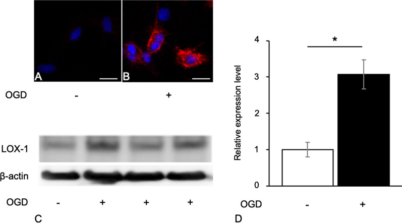
Background: Microglial cells play an important role in the immune system in the brain. Activated microglial cells are not only injurious but also neuroprotective. We confirmed marked lectin-like oxidized low-density lipoprotein receptor-1 (LOX-1) expression in microglial cells in pathological lesions in the neonatal hypoxic-ischemic encephalopathy (nHIE) model brain. LOX-1 is known to be an activator of cytokines and chemokines through intracellular pathways. Here, we investigated a novel role of LOX-1 and the molecular mechanism of LOX-1 gene transcription microglial cells under hypoxic and ischemic conditions.

Methods: We isolated primary rat microglial cells from 3-day-old rat brains and confirmed that the isolated cells showed more than 98% Iba-1 positivity with immunocytochemistry. We treated primary rat microglial cells with oxygen glucose deprivation (OGD) as an in vitro model of nHIE. Then, we evaluated the expression levels of LOX-1, cytokines and chemokines in cells treated with or without siRNA and inhibitors compared with those of cells that did not receive OGD-treatment. To confirm transcription factor binding to the OLR-1 gene promoter under the OGD conditions, we performed a luciferase reporter assay and chromatin immunoprecipitation assay. In addition, we analyzed reactive oxygen species and cell viability.

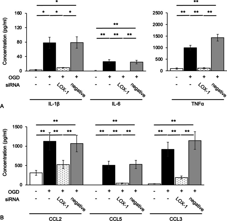
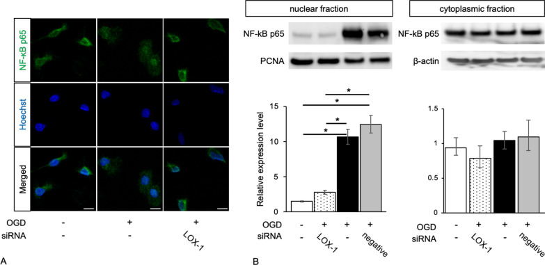
Results: We found that defects in oxygen and nutrition induced LOX-1 expression and led to the production of inflammatory mediators, such as the cytokines IL-1β, IL-6 and TNF-α; the chemokines CCL2, CCL5 and CCL3; and reactive oxygen/nitrogen species. Then, the LOX-1 signal transduction pathway was blocked by inhibitors, LOX-1 siRNA, the p38-MAPK inhibitor SB203580 and the NF-κB inhibitor BAY11-7082 suppressed the production of inflammatory mediators. We found that NF-κB and HIF-1α bind to the promoter region of the OLR-1 gene. Based on the results of the luciferase reporter assay, NF-κB has strong transcriptional activity. Moreover, we demonstrated that LOX-1 in microglial cells was autonomously overexpressed by positive feedback of the intracellular LOX-1 pathway.

Conclusion: The hypoxic/ischemic conditions of microglial cells induced LOX-1 expression and activated the immune system. LOX-1 and its related molecules or chemicals may be major therapeutic candidates. Video abstract.

Keywords: Hypoxia; Ischemia; LOX-1; Microglia; NF‐kappa B (NF‐κB); OLR-1; p38-MAPK.


Conflict of interest statement

The authors declare that they have no conflicts of interest related to the contents of this article.

Figures

Fig. 1
Fig. 1
OGD-treated microglial cells express LOX-1. Untreated microglial cells (control; CTL) did not exhibit LOX-1 expression (A). OGD-treated microglial cells (OGD) exhibited LOX-1 expression in the cytoplasm (B). Immunoblot analysis shows LOX-1 overexpression in OGD-treated microglial cells (C, D). Immunocytochemistry, blue: Hoechst 33342 (A, B), red; LOX-1 (B). *P < 0.05. Scale bar = 10 µm
Fig. 2
Fig. 2
LOX-1 siRNA inhibits OGD-induced cytokine and chemokine production. The cytokines IL-1β, IL-6 and TNF-α (A) and the chemokines CCL2, CCL5 and CCL3 (B) were significantly increased. LOX-1 siRNA downregulates the expression of these molecules. **P < 0.01
Fig. 3
Fig. 3
LOX-1 inhibition alters the distribution of NF-κB p65 from the cytoplasm to the nucleus. OGD-treated microglial cells show NF-κB p65 in the nucleus, but LOX-1 siRNA induces cytoplasmic expression of NF-κB p65 (A). Western blots exhibit significantly increased NF-κB p65 expression levels in the nuclear fraction of OGD-treated microglial cells but no significant difference in NF-κB p65 expression in the cytoplasmic fraction (B). Hoechst; Hoechst 33342, *P < 0.05. Scale bar = 10 µm
Fig. 4
Fig. 4
LOX-1 suppression induces different expression patterns of p38-MAPK and ERK phosphorylation in OGD-treated microglial cells. The expression of phospho-p38-MAPK and the ratio of phospho/total p38-MAPK were significantly increased in OGD-treated microglial cells but were reduced to the control level by LOX-1 siRNA (A). However, the expression of phospho-ERK and the ratio of phospho/total ERK were relatively increased in OGD-treated microglial cells but were not significantly reduced to the control level by LOX-1 siRNA (B). *P < 0.05
Fig. 5
Fig. 5
p38-MAPK and NF-κB inhibitors do not change LOX-1 expression levels in OGD-treated microglial cells. OGD-treated microglial cells expressed LOX-1 protein (red) in the cytoplasm (A). The same photographs of untreated microglial cells and OGD-treated microglial cells are shown in Fig. 1A, B. LOX-1 expression is not influenced by the p38-MAPK or NF-κB inhibitors SB or BAY, respectively. The relative LOX-1 expression level was the same as that shown by immunocytochemistry (B). SB; SB203580, BAY; BAY11-7082, Scale bar = 10 µm. *P < 0.05
Fig. 6
Fig. 6
The production of cytokines and chemokines in OGD-treated microglial cells is downregulated by p38-MAPK and NF-κB inhibitors. The production of the cytokines IL-1β, IL-6 and TNF-α (A) and the chemokines CCL2, CCL5 and CCL3 (B), which are increased by OGD treatment, are suppressed by SB and BAY to the same levels as those of the control. SB; SB203580, BAY; BAY11-7082, **P < 0.01
Fig. 7
Fig. 7
p38-MAPK and NF-κB inhibitors alter the distribution pattern of NF-κB p65 from the cytoplasm to the nucleus. NF-κB p65 is located in the nucleus in OGD-treated microglial cells but in the cytoplasm in other microglial cells (A). The same photographs of untreated microglial cells and OGD-treated microglial cells are shown in Fig. 3A. Western blots exhibit significantly increased NF-κB p65 expression levels in the nuclear fraction of OGD-treated microglial cells but no significant differences in NF-κB p65 expression levels in the cytoplasmic fraction (B). Hoechst; Hoechst 33,342, Scale bar = 10 µm. *P < 0.05
Fig. 8
Fig. 8
A luciferase reporter assay revealed the NF-κB binding site of − 1628 to − 1621 of the OLR-1 promoter region. The OLR-1 transcription activity is less affected by the binding sites of OCT-1 and HIF-1α. #P < 0.05 compared with the mock; formula image , not significant compared with the SV40 promoter. formula image , TATA box; formula image , NF-κB binding site; formula image , OCT-1 binding site; formula image , HIF-1α binding site; formula image , mutation position

References

    1. Ginhoux F, Greter M, Leboeuf M, Nandi S, See P, Gokhan S, et al. Fate mapping analysis reveals that adult microglia derive from primitive macrophages. Science. 2010;330:841–845. doi: 10.1126/science.1194637. - DOI - PMC - PubMed
    1. Saijo K, Glass CK. Microglial cell origin and phenotypes in health and disease. Nat Rev Immunol. 2011;11:775–787. doi: 10.1038/nri3086. - DOI - PubMed
    1. Schulz C, Gomez Perdiguero E, Chorro L, Szabo-Rogers H, Cagnard N, Kierdorf K, et al. A lineage of myeloid cells independent of Myb and hematopoietic stem cells. Science. 2012;336:86–90. doi: 10.1126/science.1219179. - DOI - PubMed
    1. Faustino JV, Wang X, Johnson CE, Klibanov A, Derugin N, Wendland MF, et al. Microglial cells contribute to endogenous brain defenses after acute neonatal focal stroke. J Neurosci. 2011;31:12992–13001. doi: 10.1523/JNEUROSCI.2102-11.2011. - DOI - PMC - PubMed
    1. Jin WN, Shi SX, Li Z, Li M, Wood K, Gonzales RJ, et al. Depletion of microglia exacerbates postischemic inflammation and brain injury. J Cereb Blood Flow Metab. 2017;37:2224–2236. doi: 10.1177/0271678X17694185. - DOI - PMC - PubMed

Publication types

LinkOut - more resources