. 2023 Jun 3;21(1):361.
doi: 10.1186/s12967-023-04198-0.

Dental pulp stem cell-derived exosomes revitalize salivary gland epithelial cell function in NOD mice via the GPER-mediated cAMP/PKA/CREB signaling pathway

Affiliations

Dental pulp stem cell-derived exosomes revitalize salivary gland epithelial cell function in NOD mice via the GPER-mediated cAMP/PKA/CREB signaling pathway

Shilin Hu et al. J Transl Med. .

Abstract

Background: Restoration of salivary gland function in Sjogren's syndrome (SS) is still a challenge. Dental pulp stem cells (DPSCs) derived exosomes had shown anti-inflammatory, anti-oxidative, immunomodulatory, and tissue function restorative abilities. However, the salivary gland function restoration potential of DPSCs-derived exosomes (DPSC-Exos) during SS has not been investigated yet.

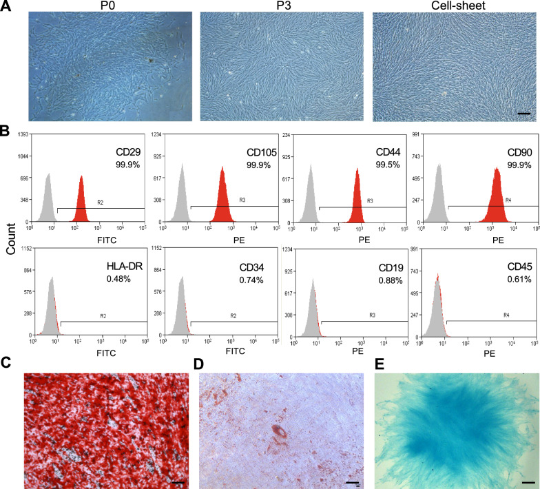
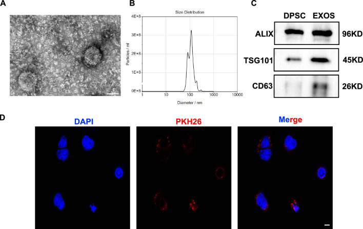
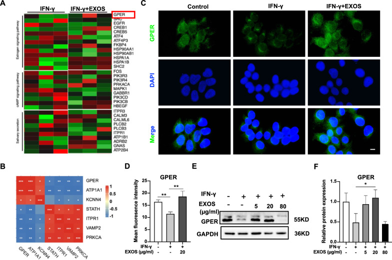
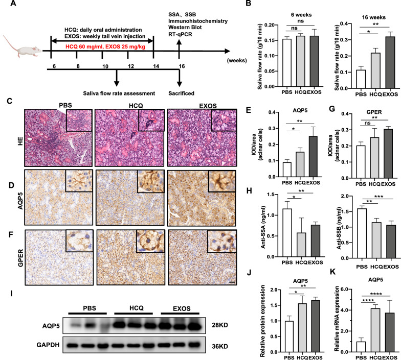
Methods: DPSC-Exos was isolated by ultracentrifugation methods and characterized. Salivary gland epithelial cells (SGEC) were treated with interferon-gamma (IFN-γ) to mimic SS in vitro and cultured with or without DPSC-Exos. SGEC survival and aquaporin 5 (AQP5) expression were analyzed. mRNA sequencing and bioinformatics analysis were performed in IFN-γ vs. DPSC-Exos+ IFN-γ treated SGEC. Non-obese diabetic (NOD)/ltj female mice (SS model), were intravenously administered with DPSC-Exos, and salivary gland functions and SS pathogenicity were analyzed. Furthermore, the mRNA sequencing and bioinformatics predicted mechanism of the therapeutic effect of DPSC-Exos was further investigated both in vitro and in vivo using RT-qPCR, Western blot, immunohistochemistry, immunofluorescence, flowcytometry analysis.

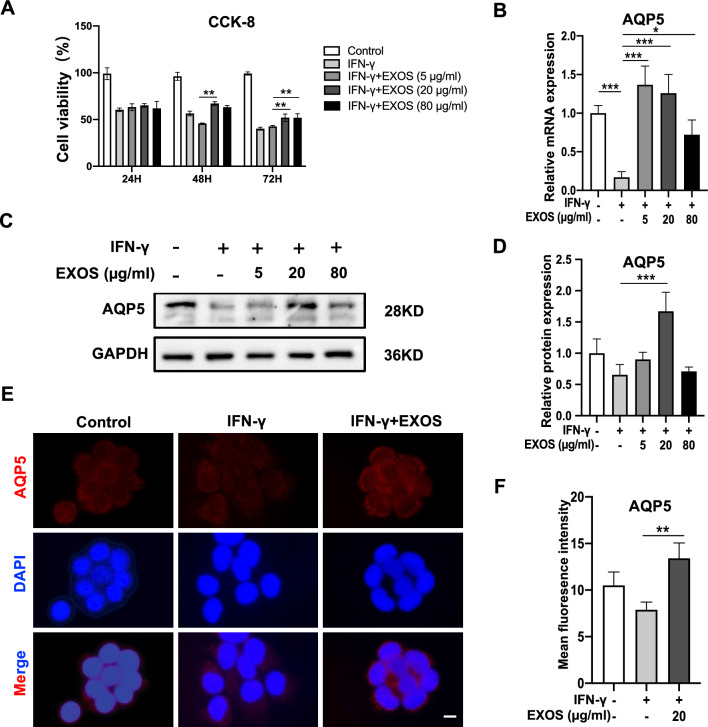
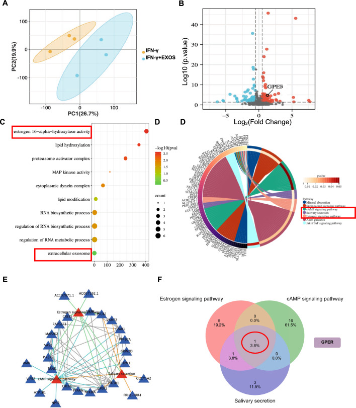
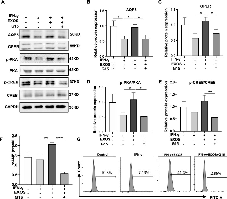
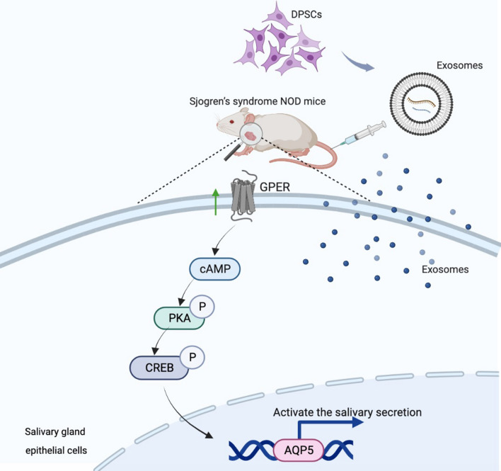
Results: DPSC-Exos partially rescued IFN-γ triggered SGEC death. IFN-γ inhibited AQP5 expression in SGEC and DPSC-Exos reversed this effect. Transcriptome analysis showed GPER was the upregulated DEG in DPSC-Exos-treated SGEC with a positive correlation with salivary secretion-related DEGs. Pathway enrichment analysis revealed that DEGs were mainly attributed to estrogen 16 alpha-hydroxylase activity, extracellular exosome function, cAMP signaling, salivary secretion, and estrogen signaling. Intravenous injection of DPSC-Exos in NOD/ltj mice alleviated the SS syndrome as indicated by the increased salivary flow rate, attenuated glandular inflammation, and increased AQP5 expression. GPER was also upregulated in the salivary gland of DPSC-Exos-treated NOD/ltj mice compared with the PBS-treated NOD/ltj mice. IFN-γ+DPSC-Exos-treated SGEC showed higher expression of AQP5, p-PKA, cAMP, and intracellular Ca2+ levels compared with IFN-γ-treated SGEC. These effects were reversed by the inhibition of GPER.

Conclusions: Our results showed that DPSC-Exos revitalize salivary gland epithelial cell function during SS via the GPER-mediated cAMP/PKA/CREB pathway suggesting the possible therapeutic potential of DPSC-Exos in SS-treatment.

Keywords: Aquaporin 5 (AQP5); Dental pulp stem cells (DPSC); Exosomes; G-protein coupled estrogen receptor (GPER); Salivary gland epithelial cells (SGEC); Sjogren’s syndrome (SS).


Conflict of interest statement

The authors declare that they have no competing interests.

Figures

Fig. 1
Fig. 1
Isolation and characterization of DPSCs. A Human DPSCs culture at primary passage (P0), passage 3 (P3), and cell sheet. Scale bar: 200 μm. B Analysis of DPSCs surface markers by flow cytometry. C Alizarin red, D oil red O, and E alcian blue staining of DPSCs after inducing osteogenic, adipogenic, and chondrogenic differentiation respectively. Scale bar: 100 μm
Fig. 2
Fig. 2
Isolation and characterization of DPSCs cell sheet released exosomes-released exosomes (DPSC-Exos). A Representative TEM images of DPSC-Exos. Scale bar: 100 nm. B Nanoparticle tracking analysis of DPSC-Exos. C Western blot analysis of exosome-specific markers ALIX, TSG101, and CD6. D Immunofluorescence staining for DPSC-Exos uptake by salivary gland epithelial cells (SGEC). Scale bar: 20 μm
Fig. 3
Fig. 3
DPSC-Exos rescued IFN-γ-caused death and AQP5 downregulation of salivary gland epithelial cells (SGEC). A The cell viability of salivary gland epithelial cells was detected by CCK-8 assay. The expression pattern of AQP5 analyzed by RT-qPCR (B), Western blot analysis (C, D), and immunofluorescence staining (E, F). Scale bar: 20 μm. Data are presented as the mean ± SD, n = 3. Significant effect of the treatment, *p < 0.05, **p < 0.01, ***p < 0.001
Fig. 4
Fig. 4
Transcriptome analysis of salivary gland epithelial cells treated with or without DPSC-Exos. A The Principal Component Analysis (PCA). B Volcano plot showing differentially expressed genes (DEGs). C Gene ontology analysis. D KEGG analysis enriched GPER-related pathways among DEGs. E STRING database shows the interaction among the cAMP signaling pathway, salivary secretion, and estrogen signaling pathway, and F Venn analysis of the genes among the cAMP signaling pathway, salivary secretion, and estrogen signaling pathway. All DEGs are screened based on P-value < 0.05 and |fold change| > 1
Fig. 5
Fig. 5
DPSC-Exos treatment induced GPER expression in salivary gland epithelial cells (SGEC). A Heatmap of differentially expressed genes (DEGs) related to cAMP signaling pathway, salivary secretion, and estrogen signaling pathway. B Correlation heatmap between GPER and the marker of salivary secretion in DEGs. C, D Immunofluorescence staining for GPER expression in SGEC. E, F Western blot analysis of GPER expression in SGEC. Scale bar: 20 μm. Data are presented as the mean ± SD, n = 3. Significant effect of the treatment, *p < 0.05, **p < 0.01, ***p < 0.001
Fig. 6
Fig. 6
DPSC-Exos treatment alleviated SS-like symptoms in NOD/ltj mice. A Scheme of animal experiment and treatment. B The saliva flow rate, C H&E stained histological images of mice submandibular glands. Immunohistochemistry analysis of AQP5 (D, E) and GPER (F, G) expression in mice submandibular glands. H The anti-SSA/SSB levels of NOD/ltj mice serum were detected by ELISA. Western blot (I, J) and RT-qPCR analysis (K) of AQP5 expression in mice submandibular. Scale bar: 100 μm. Data are presented as the mean ± SD, n = 6. Significant effect of the treatment, *p < 0.05, **p < 0.01, ***p < 0.001
Fig. 7
Fig. 7
DPSC-Exos alleviated IFN-γ-caused AQP5 downregulation in salivary gland epithelial cells (SGEC) via GPER mediated cAMP-PKA-CREB pathway. AE Western blot analysis AQP5, GPER, p-PKA, and p-CREB in SGEC. F The cAMP concentration in SGEC lysate was detected by ELISA. G The intracellular Ca2+ level of SGEC was measured by flow cytometry. Data are presented as the mean ± SD, n = 3. Significant effect of the treatment, *p < 0.05, **p < 0.01, ***p < 0.001
Fig. 8
Fig. 8
Schematic diagram of DPSC-Exos-mediated activation of saliva secretory function in NOD/ltj mice

References

    1. Le Gall M, Cornec D, Pers JO, Saraux A, Jousse-Joulin S, Cochener B, et al. A prospective evaluation of dental and periodontal status in patients with suspected Sjögren’s syndrome. Joint Bone Spine. 2016;83(2):235–236. doi: 10.1016/j.jbspin.2015.02.015. - DOI - PubMed
    1. Sarkar I, Davies R, Aarebrot AK, Solberg SM, Petrovic A, Joshi AM, et al. Aberrant signaling of immune cells in Sjögren’s syndrome patient subgroups upon interferon stimulation. Front Immunol. 2022;13:854183. doi: 10.3389/fimmu.2022.854183. - DOI - PMC - PubMed
    1. Vivino FB, Bunya VY, Massaro-Giordano G, Johr CR, Giattino SL, Schorpion A, et al. Sjogren’s syndrome: an update on disease pathogenesis, clinical manifestations and treatment. Clin Immunol. 2019;203:81–121. doi: 10.1016/j.clim.2019.04.009. - DOI - PubMed
    1. Salum FG, Medella-Junior FDAC, Figueiredo MAZ, Cherubini K. Salivary hypofunction: an update on therapeutic strategies. Gerodontology. 2018;35(4):305–316. doi: 10.1111/ger.12353. - DOI - PubMed
    1. Lai Z, Yin H, Cabrera-Pérez J, Guimaro MC, Afione S, Michael DG, et al. Aquaporin gene therapy corrects Sjögren’s syndrome phenotype in mice. Proc Natl Acad Sci USA. 2016;113(20):5694–5699. doi: 10.1073/pnas.1601992113. - DOI - PMC - PubMed

Publication types