. 2023 Jun 2;17(1):45.
doi: 10.1186/s40246-023-00490-8.

Long-read genome sequencing identifies cryptic structural variants in congenital aniridia cases

Affiliations

Long-read genome sequencing identifies cryptic structural variants in congenital aniridia cases

Alejandra Damián et al. Hum Genomics. .

Abstract

Background: Haploinsufficiency of the transcription factor PAX6 is the main cause of congenital aniridia, a genetic disorder characterized by iris and foveal hypoplasia. 11p13 microdeletions altering PAX6 or its downstream regulatory region (DRR) are present in about 25% of patients; however, only a few complex rearrangements have been described to date. Here, we performed nanopore-based whole-genome sequencing to assess the presence of cryptic structural variants (SVs) on the only two unsolved "PAX6-negative" cases from a cohort of 110 patients with congenital aniridia after unsuccessfully short-read sequencing approaches.

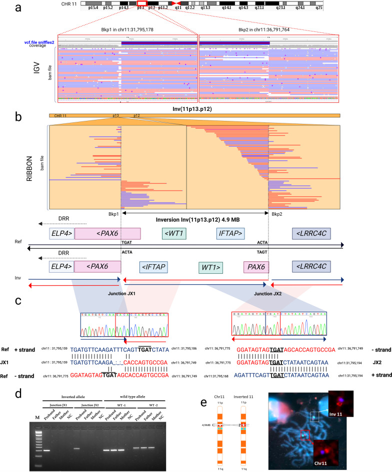
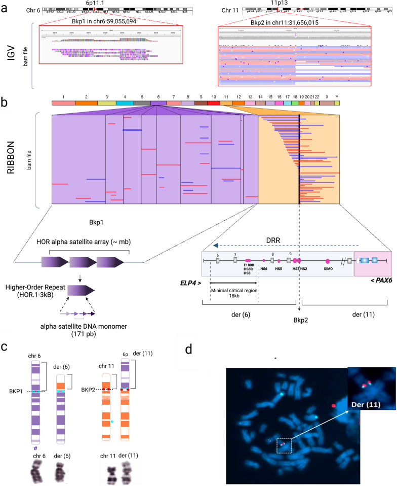
Results: Long-read sequencing (LRS) unveiled balanced chromosomal rearrangements affecting the PAX6 locus at 11p13 in these two patients and allowed nucleotide-level breakpoint analysis. First, we identified a cryptic 4.9 Mb de novo inversion disrupting intron 7 of PAX6, further verified by targeted polymerase chain reaction amplification and sequencing and FISH-based cytogenetic analysis. Furthermore, LRS was decisive in correctly mapping a t(6;11) balanced translocation cytogenetically detected in a second proband with congenital aniridia and considered non-causal 15 years ago. LRS resolved that the breakpoint on chromosome 11 was indeed located at 11p13, disrupting the DNase I hypersensitive site 2 enhancer within the DRR of PAX6, 161 Kb from the causal gene. Patient-derived RNA expression analysis demonstrated PAX6 haploinsufficiency, thus supporting that the 11p13 breakpoint led to a positional effect by cleaving crucial enhancers for PAX6 transactivation. LRS analysis was also critical for mapping the exact breakpoint on chromosome 6 to the highly repetitive centromeric region at 6p11.1.

Conclusions: In both cases, the LRS-based identified SVs have been deemed the hidden pathogenic cause of congenital aniridia. Our study underscores the limitations of traditional short-read sequencing in uncovering pathogenic SVs affecting low-complexity regions of the genome and the value of LRS in providing insight into hidden sources of variation in rare genetic diseases.

Keywords: Aniridia; Chromosomal rearrangements; Long-read genome sequencing; Nanopore sequencing; PAX6.


Conflict of interest statement

The authors declare no competing interests.

Figures

Fig. 1
Fig. 1
Identification and validation of a balanced paracentric inversion involving PAX6 in patient ANI-1. a–b. Representation of long reads, breakpoints, and genomic elements involved in a 4.9 Mb paracentric inversion on chromosome 11. Nanopore long-read genome sequencing from high molecular weight DNA revealed a cryptic heterozygous inversion disrupting PAX6. The IGV and Ribbon tools were used to visualize read alignments from vcf and BAM files. Genes and regulatory elements most relevant to phenotypic expression of congenital aniridia, including PAX6 and downstream regulatory regions (DRR) and WT1 and adjacent genes next to the breakpoints, are depicted. BKP1 and BKP2: distal and proximal breakpoints. c. Validation of the distal breakpoint of the inverted allele fragment junctions (JX1 and JX2) on both sides by PCR and Sanger sequencing. d. Electrophoresis of the amplified-fragment junctions in the proband showed unique bands corresponding to the inverted allele in JX1 and JX2, while amplicons for the wild-type allele were also present in both parents. NTC: no template control. e. Schematic representation of the inversion and FISH findings. Centromeric D11Z1 and PAX6 probes are labeled in cyan blue and red, respectively. FISH analysis showed a well-defined red-associated PAX6 signal on wild-type chromosome 11 and spread and separated red-associated PAX6 signals on the inverted chromosome
Fig. 2
Fig. 2
Identification of a reciprocal translocation t(6;11) affecting the PAX6 downstream regulatory region in patient ANI-2. a–b. Representation of long-reads, breakpoints, and genomic elements involved in the paracentric inversion using IGV and Ribbon visualizers from vcf and BAM files. LRS showed evidence of an interchromosomal translocation involving alpha-satellite DNA sequences from the centromeric region of chromosome 6 (6p11.1) and their mate split reads laying on chromosome 11 (11p13) in intron 9 of the ELP4 gene. The breakpoint on 11p13 disrupts the DNase I hypersensitive site 2 (HS2) enhancer within the downstream regulatory region (DRR) of PAX6. This chromosomal break is taking apart PAX6 from its critical 18 Kb minimal regulatory region defined by Plaisancie et al., 2018 [10]. c–d. Karyotyping and FISH analysis showed the presence of a t(6;11) reciprocal translocation. Schematic representation of the FISH assay designed to visualize the breakpoints. The centromeric CEP6-D6Z1 and PAX6 probes are labeled in cyan blue and red, respectively. FISH images showed specific signals for PAX6 and CEP6-D6Z1 alpha-satellite sequences on derivative chromosome 11, der (11). A signal for CEP6-D6Z1 was also observed on derivative chromosome 6, der(6), confirming breakpoint involvement at the centromeric level
Fig. 3
Fig. 3
Functional characterization of SVs affecting PAX6 expression. Boxplots represent the relative PAX6 mRNA expression in a lymphocyte cell line derived from both SV-carrying probands compared to three healthy controls (wt-PAX6) and three patients carrying PAX6-associated aniridia pathogenic variants from our cohort. RPLP0 gene was used for setting relative expression. The experiment was performed in triplicate. Boxplots indicate median values and range of whiskers variation. FC: Fold-change indicating the normalized ratio of PAX6/RPLP0 expression levels. Statistical analysis was performed using Kruskal–Wallis test with Dunn's test for multiple comparisons of normalized PAX6 expression ratios and Bonferroni for multiple testing corrections. Significance was defined as **p.adj < 0.01, ****p.adj < 0.0001, and p.adj > 0.05 (not significant, ns)

References

    1. Hingorani M, Hanson I, van Heyningen V. Aniridia. Eur J Hum Genet. 2012;20:1011–1017. doi: 10.1038/ejhg.2012.100. - DOI - PMC - PubMed
    1. Lima Cunha D, Arno G, Corton M, Moosajee M. The spectrum of PAX6 mutations and genotype-phenotype correlations in the eye. Genes. 2019;10:1050. doi: 10.3390/genes10121050. - DOI - PMC - PubMed
    1. Cvekl A, Callaerts P. PAX6: 25th anniversary and more to learn. Exp Eye Res. 2017;156:10–21. doi: 10.1016/j.exer.2016.04.017. - DOI - PubMed
    1. Bhatia S, Monahan J, Ravi V, Gautier P, Murdoch E, Brenner S, et al. A survey of ancient conserved non-coding elements in the PAX6 locus reveals a landscape of interdigitated cis-regulatory archipelagos. Dev Biol. 2014;387:214–228. doi: 10.1016/j.ydbio.2014.01.007. - DOI - PubMed
    1. Blanco-Kelly F, Palomares M, Vallespín E, Villaverde C, Martín-Arenas R, Vélez-Monsalve C, et al. Improving molecular diagnosis of aniridia and WAGR syndrome using customized targeted array-based CGH. PLoS ONE. 2017;12:e0172363. doi: 10.1371/journal.pone.0172363. - DOI - PMC - PubMed

Publication types

Substances

LinkOut - more resources