

Molecular pathogen screening of louse flies (Diptera: Hippoboscidae) from domestic and wild ruminants in Austria
- PMID: 37269018
- PMCID: PMC10236838
- DOI: 10.1186/s13071-023-05810-4
Molecular pathogen screening of louse flies (Diptera: Hippoboscidae) from domestic and wild ruminants in Austria
Abstract
Background: Hippoboscid flies (Diptera: Hippoboscidae), also known as louse flies or keds, are obligate blood-sucking ectoparasites of animals, and accidentally of humans. The potential role of hippoboscids as vectors of human and veterinary pathogens is being increasingly investigated, but the presence and distribution of infectious agents in louse flies is still unknown in parts of Europe. Here, we report the use of molecular genetics to detect and characterize vector-borne pathogens in hippoboscid flies infesting domestic and wild animals in Austria.
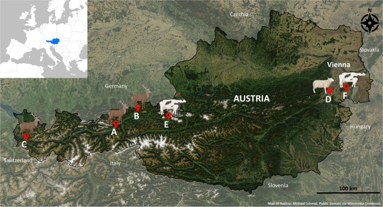
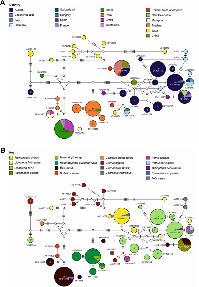
Methods: Louse flies were collected from naturally infested cattle (n = 25), sheep (n = 3), and red deer (n = 12) across Austria between 2015 and 2019. Individual insects were morphologically identified to species level and subjected to DNA extraction for molecular pathogen screening and barcoding. Genomic DNA from each louse fly was screened for Borrelia spp., Bartonella spp., Trypanosomatida, Anaplasmataceae, Filarioidea and Piroplasmida. Obtained sequences of Trypanosomatida and Bartonella spp. were further characterized by phylogenetic and haplotype networking analyses.
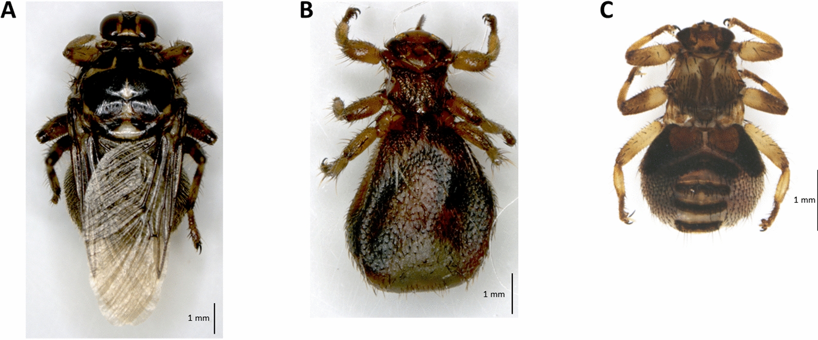
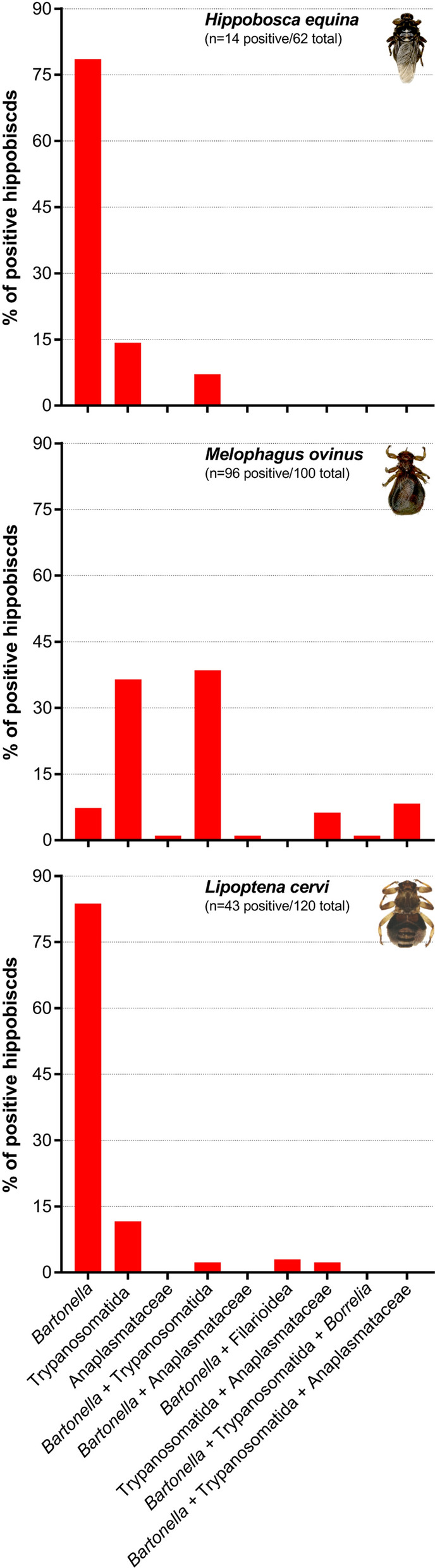
Results: A total of 282 hippoboscid flies corresponding to three species were identified: Hippobosca equina (n = 62) collected from cattle, Melophagus ovinus (n = 100) from sheep and Lipoptena cervi (n = 120) from red deer (Cervus elaphus). Molecular screening revealed pathogen DNA in 54.3% of hippoboscids, including infections with single (63.39%), two (30.71%) and up to three (5.90%) distinct pathogens in the same individual. Bartonella DNA was detected in 36.9% of the louse flies. Lipoptena cervi were infected with 10 distinct and previously unreported Bartonella sp. haplotypes, some closely associated with strains of zoonotic potential. DNA of trypanosomatids was identified in 34% of hippoboscids, including the first description of Trypanosoma sp. in H. equina. Anaplasmataceae DNA (Wolbachia spp.) was detected only in M. ovinus (16%), while < 1% of the louse flies were positive for Borrelia spp. and Filarioidea. All hippoboscids were negative for Piroplasmida.
Conclusions: Molecular genetic screening confirmed the presence of several pathogens in hippoboscids infesting domestic and wild ruminants in Austria, including novel pathogen haplotypes of zoonotic potential (e.g. Bartonella spp.) and the first report of Trypanosoma sp. in H. equina, suggesting a potential role of this louse fly as vector of animal trypanosomatids. Experimental transmission studies and expanded monitoring of hippoboscid flies and hippoboscid-associated pathogens are warranted to clarify the competence of these ectoparasites as vectors of infectious agents in a One-Health context.
Keywords: Barcoding; Bartonella; Hippobosca equina; Hippoboscidae; Keds; Lipoptena cervi; Louse flies; Melophagus ovinus; Ruminants; Vector-borne pathogens.
© 2023. The Author(s).
Conflict of interest statement
The authors declare no competing interests.
Figures

Similar articles
-
Detection of Bartonella schoenbuchensis (sub)species DNA in different louse fly species in Saxony, Germany: The proof of multiple PCR analysis necessity in case of ruminant-associated bartonellae determination.Vet Med Sci. 2024 May;10(3):e1417. doi: 10.1002/vms3.1417. Vet Med Sci. 2024. PMID: 38516829 Free PMC article.
-
Molecular evidence of bacteria in Melophagus ovinus sheep keds and Hippobosca equina forest flies collected from sheep and horses in northeastern Algeria.Comp Immunol Microbiol Infect Dis. 2019 Aug;65:103-109. doi: 10.1016/j.cimid.2019.05.010. Epub 2019 May 10. Comp Immunol Microbiol Infect Dis. 2019. PMID: 31300097
-
Molecular and morphological analysis revealed a new Lipoptena species (Diptera: Hippoboscidae) in southern Spain harbouring Coxiella burnetii and bacterial endosymbionts.Vet Parasitol. 2024 Dec;332:110300. doi: 10.1016/j.vetpar.2024.110300. Epub 2024 Sep 3. Vet Parasitol. 2024. PMID: 39270602
-
Keds, the enigmatic flies and their role as vectors of pathogens.Acta Trop. 2020 Sep;209:105521. doi: 10.1016/j.actatropica.2020.105521. Epub 2020 May 21. Acta Trop. 2020. PMID: 32447028 Review.
-
A brief review on deer keds of the genus Lipoptena (Diptera: Hippoboscidae).Vet Parasitol. 2023 Jan;313:109850. doi: 10.1016/j.vetpar.2022.109850. Epub 2022 Dec 2. Vet Parasitol. 2023. PMID: 36473321 Review.
Cited by
-
A new species of louse fly, Ornithomya Latreille, 1802 (Diptera: Hippoboscidae), from the Russian far East.Int J Parasitol Parasites Wildl. 2025 Jul 1;27:101111. doi: 10.1016/j.ijppaw.2025.101111. eCollection 2025 Aug. Int J Parasitol Parasites Wildl. 2025. PMID: 40697857 Free PMC article.
-
Chewing lice (Phthiraptera) on a wild Golden eagle (Aquila chrysaetos) and a zoo-kept Eurasian griffon vulture (Gyps fulvus) in Tyrol, Austria.Parasitol Res. 2025 Jul 21;124(7):85. doi: 10.1007/s00436-025-08531-y. Parasitol Res. 2025. PMID: 40685410 Free PMC article.
-
Global Mapping and Visualization Analysis of One Health Knowledge in the COVID-19 Context.Environ Health Insights. 2024 Mar 5;18:11786302241236017. doi: 10.1177/11786302241236017. eCollection 2024. Environ Health Insights. 2024. PMID: 38449589 Free PMC article.
-
Louse flies (Diptera: Hippoboscidae) of Romania: New records and novel host-parasite and hyperparasites associations.Int J Parasitol Parasites Wildl. 2025 Jun 6;27:101100. doi: 10.1016/j.ijppaw.2025.101100. eCollection 2025 Aug. Int J Parasitol Parasites Wildl. 2025. PMID: 40547399 Free PMC article.
-
Vector-borne pathogens in dogs from the Republic of Kosovo.Parasit Vectors. 2025 Apr 9;18(1):136. doi: 10.1186/s13071-025-06777-0. Parasit Vectors. 2025. PMID: 40205569 Free PMC article.
References
-
- Reeves WK, Lloyd JE. Chapter20: louse flies, keds, and bat flies (Hippoboscoidea) In: Mullen GR, Durden LA, editors. Medical and veterinary entomology. Amsterdam: Elsevier Inc; 2019. pp. 421–38.
-
- Smart J. Ked-flies. Nature. 1945;155:123. doi: 10.1038/155123a0. - DOI
-
- Rantanen T, Reunala T, Vuojolahti P, Hackman W. Persistent pruritic papules from deer ked bites. Acta Derm Venereol. 1982;62:307–311. - PubMed
MeSH terms
Grants and funding
LinkOut - more resources
Full Text Sources
Miscellaneous
