

TNF-α induced extracellular release of keratinocyte high-mobility group box 1 in Stevens-Johnson syndrome/toxic epidermal necrolysis: Biomarker and putative mechanism of pathogenesis
- PMID: 37269158
- PMCID: PMC10947163
- DOI: 10.1111/1346-8138.16847
TNF-α induced extracellular release of keratinocyte high-mobility group box 1 in Stevens-Johnson syndrome/toxic epidermal necrolysis: Biomarker and putative mechanism of pathogenesis
Abstract
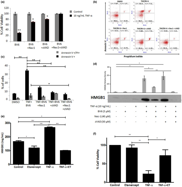
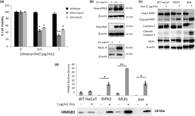
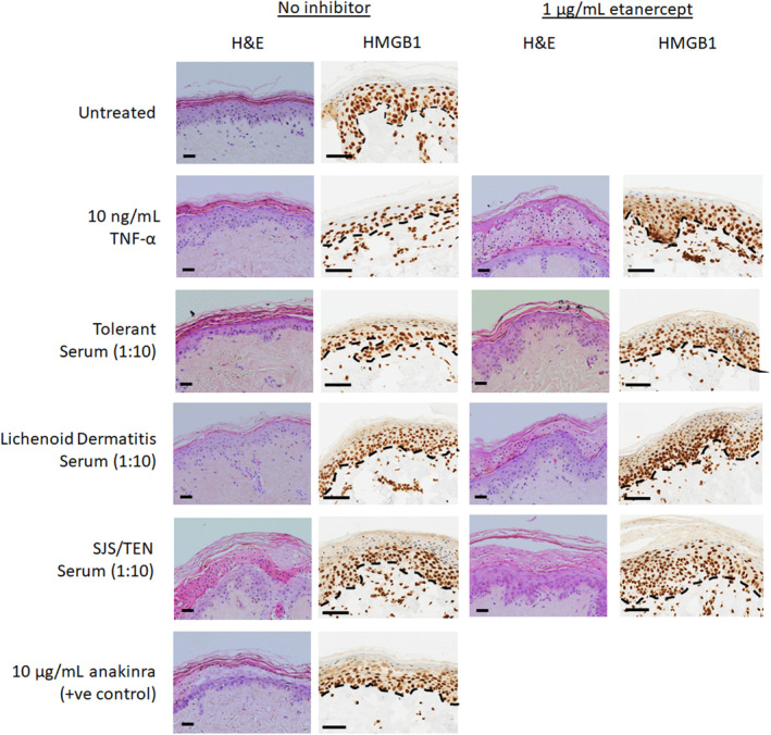
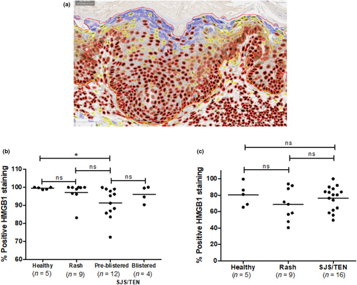
Decreased epidermal high-mobility group box 1 (HMGB1) expression is an early marker of epidermal injury in Stevens-Johnson syndrome/toxic epidermal necrolysis (SJS/TEN). Etanercept, an anti-tumor necrosis factor therapeutic, is effective in the treatment of SJS/TEN. The objective was to characterize antitumor necrosis factor-alpha (TNF-α)-mediated HMGB1 keratinocyte/epidermal release and etanercept modulation. HMGB1 release from TNF-α treated (± etanercept), or doxycycline-inducible RIPK3 or Bak-expressing human keratinocyte cells (HaCaTs) was determined by western blot/ELISA. Healthy skin explants were treated with TNF-α or serum (1:10 dilution) from immune checkpoint inhibitor-tolerant, lichenoid dermatitis or SJS/TEN patients ± etanercept. Histological and immunohistochemical analysis of HMGB1 was undertaken. TNF-α induced HMGB1 release in vitro via both necroptosis and apoptosis. Exposure of skin explants to TNF-α or SJS/TEN serum resulted in significant epidermal toxicity/detachment with substantial HMGB1 release which was attenuated by etanercept. Whole-slide image analysis of biopsies demonstrated significantly lower epidermal HMGB1 in pre-blistered SJS/TEN versus control (P < 0.05). Keratinocyte HMGB1 release, predominantly caused by necroptosis, can be attenuated by etanercept. Although TNF-α is a key mediator of epidermal HMGB1 release, other cytokines/cytotoxic proteins also contribute. Skin explant models represent a potential model of SJS/TEN that could be utilized for further mechanistic studies and targeted therapy screening.
Keywords: Stevens-Johnson syndrome; high-mobility group box 1; toxic epidermal necrolysis; tumor necrosis factor alpha.
© 2023 The Authors. The Journal of Dermatology published by John Wiley & Sons Australia, Ltd on behalf of Japanese Dermatological Association.
Conflict of interest statement
Etanercept (Benepali) was donated to DFC by Biogen Inc. The authors report no other conflicts of interest.
Figures
Similar articles
-
TNF-α‒Mediated Keratinocyte Expression and Release of Matrix Metalloproteinase 9: Putative Mechanism of Pathogenesis in Stevens‒Johnson Syndrome/Toxic Epidermal Necrolysis.J Invest Dermatol. 2023 Jun;143(6):1023-1030.e7. doi: 10.1016/j.jid.2022.11.024. Epub 2022 Dec 26. J Invest Dermatol. 2023. PMID: 36581093
-
Serum and blister-fluid elevation and decreased epidermal content of high-mobility group box 1 protein in drug-induced Stevens-Johnson syndrome/toxic epidermal necrolysis.Br J Dermatol. 2019 Jul;181(1):166-174. doi: 10.1111/bjd.17610. Epub 2019 Mar 26. Br J Dermatol. 2019. PMID: 30613954 Free PMC article.
-
Evaluation of Combination Therapy With Etanercept and Systemic Corticosteroids for Stevens-Johnson Syndrome and Toxic Epidermal Necrolysis: A Multicenter Observational Study.J Allergy Clin Immunol Pract. 2022 May;10(5):1295-1304.e6. doi: 10.1016/j.jaip.2022.01.038. Epub 2022 Feb 4. J Allergy Clin Immunol Pract. 2022. PMID: 35131514
-
Recent advances in managing and understanding Stevens-Johnson syndrome and toxic epidermal necrolysis.F1000Res. 2020 Jun 16;9:F1000 Faculty Rev-612. doi: 10.12688/f1000research.24748.1. eCollection 2020. F1000Res. 2020. PMID: 32595945 Free PMC article. Review.
-
Biologic TNF-alpha inhibitors in the treatment of Stevens-Johnson syndrome and toxic epidermal necrolysis: a systemic review.J Dermatolog Treat. 2020 Feb;31(1):66-73. doi: 10.1080/09546634.2019.1577548. Epub 2019 Feb 19. J Dermatolog Treat. 2020. PMID: 30702955
Cited by
-
Update on Stevens-Johnson Syndrome and Toxic Epidermal Necrolysis: Diagnosis and Management.Am J Clin Dermatol. 2024 Nov;25(6):891-908. doi: 10.1007/s40257-024-00889-6. Epub 2024 Sep 15. Am J Clin Dermatol. 2024. PMID: 39278968 Free PMC article. Review.
-
Evaluating the Combined Effect of a Choline Kinase Inhibitor and Temozolomide Therapy in a Mouse Model of Glioblastoma Using 1H MR Spectroscopy and IVIM-DWI.NMR Biomed. 2025 Sep;38(9):e70113. doi: 10.1002/nbm.70113. NMR Biomed. 2025. PMID: 40755153 Free PMC article.
-
An In Vitro Human Skin Test for Predicting Skin Sensitization and Adverse Immune Reactions to Biologics.Toxics. 2024 May 30;12(6):401. doi: 10.3390/toxics12060401. Toxics. 2024. PMID: 38922081 Free PMC article.
-
Greater mechanistic understanding of the cutaneous pathogenesis of Stevens-Johnson syndrome/toxic epidermal necrolysis can shed light on novel therapeutic strategies: a comprehensive review.Curr Opin Allergy Clin Immunol. 2024 Aug 1;24(4):218-227. doi: 10.1097/ACI.0000000000000993. Epub 2024 May 17. Curr Opin Allergy Clin Immunol. 2024. PMID: 38753537 Free PMC article. Review.
-
HMGB1: a double-edged sword and therapeutic target in the female reproductive system.Front Immunol. 2023 Aug 18;14:1238785. doi: 10.3389/fimmu.2023.1238785. eCollection 2023. Front Immunol. 2023. PMID: 37691930 Free PMC article. Review.
References
-
- Andersson U, Antoine DJ, Tracey KJ. The functions of HMGB1 depend on molecular localization and post‐translational modifications. J Intern Med. 2014;276:420–4. - PubMed
-
- Zhang S, Tang S, Li S, Pan Y, Ding Y. Biologic TNF‐alpha inhibitors in the treatment of Stevens‐Johnson syndrome and toxic epidermal necrolysis: a systemic review. J Dermatolog Treat. 2020;31:66–73. - PubMed
-
- Lee Y‐Y, Ko J‐H, Wei C‐H, Chung W‐H. Use of etanercept to treat toxic epidermal necrolysis in a human immunodeficiency virus‐positive patient. Dermatol Sin. 2013;31:78–81.
MeSH terms
Substances
Grants and funding
LinkOut - more resources
Full Text Sources
Miscellaneous
