Review
. 2023 Jun 3;15(1):145.
doi: 10.1007/s40820-023-01125-2.

Application of Nano-Delivery Systems in Lymph Nodes for Tumor Immunotherapy

Affiliations
Review

Application of Nano-Delivery Systems in Lymph Nodes for Tumor Immunotherapy

Yiming Xia et al. Nanomicro Lett. .

Abstract

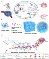
Immunotherapy has become a promising research "hotspot" in cancer treatment. "Soldier" immune cells are not uniform throughout the body; they accumulate mostly in the immune organs such as the spleen and lymph nodes (LNs), etc. The unique structure of LNs provides the microenvironment suitable for the survival, activation, and proliferation of multiple types of immune cells. LNs play an important role in both the initiation of adaptive immunity and the generation of durable anti-tumor responses. Antigens taken up by antigen-presenting cells in peripheral tissues need to migrate with lymphatic fluid to LNs to activate the lymphocytes therein. Meanwhile, the accumulation and retaining of many immune functional compounds in LNs enhance their efficacy significantly. Therefore, LNs have become a key target for tumor immunotherapy. Unfortunately, the nonspecific distribution of the immune drugs in vivo greatly limits the activation and proliferation of immune cells, which leads to unsatisfactory anti-tumor effects. The efficient nano-delivery system to LNs is an effective strategy to maximize the efficacy of immune drugs. Nano-delivery systems have shown beneficial in improving biodistribution and enhancing accumulation in lymphoid tissues, exhibiting powerful and promising prospects for achieving effective delivery to LNs. Herein, the physiological structure and the delivery barriers of LNs were summarized and the factors affecting LNs accumulation were discussed thoroughly. Moreover, developments in nano-delivery systems were reviewed and the transformation prospects of LNs targeting nanocarriers were summarized and discussed.

Keywords: Cancer therapy; Immunotherapy; Lymph nodes; Nano-delivery systems.


Conflict of interest statement

The authors declare no interest conflict. They have no known competing financial interests or personal relationships that could have appeared to influence the work reported in this paper.

Figures

Fig. 1
Fig. 1
Schematic diagram of LNs distribution, structure and barriers. Created with BioRender.com
Fig. 2
Fig. 2
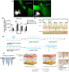
Factors affecting LNs accumulation of nanoparticles. a Schematic representation of the entry of particles of different particle sizes into the LNs [42]. Copyright 2017 Elsevier. b CLSM images of LNs treated with LNPs with positive and negative charges. LNPs with negative charges infiltrated into the inner area of the LNs [68]. Copyright 2020 American Chem. Society. c Schematic illustration of the deformable strategy of lymph-node transfer [83]. Copyright 2021 John Wiley and Sons. d The percentages (left) and MFI (right) of fluorescein-labeled naked-, mono-mannosylated- (MN), and tri-mannosylated (triMN) LPR formulations incubated with DC 2.4 cells. The triMN-LPR were significantly better associated with DC 2.4 cells than MN-LPR [93]. Copyright 2018 Elsevier. e Schematic illustration of nanocomplex-decorated microbubbles targeting CD11b on APCs [94]. Copyright 2022 Springer Nature
Fig. 3
Fig. 3
Summary of nano-delivery systems in LNs for tumor immunotherapy. a [102], Copyright 2018 American Chemical Society; b [103], Copyright 2021 American Chemical Society. c [104], Copyright 2019 American Chemical Society; d [93], Copyright 2018 Elsevier; e [105], Copyright 2022 Elsevier. f [106], Copyright 2018 Elsevier; g [107], Copyright 2021 American Chemical Society; h [108], Copyright 2022 Taylor & Francis Journal. i [109], Copyright 2021 John Wiley and Sons; j [110], Copyright 2019 Elsevier; k [111], Copyright 2021 Multidisciplinary Digital Publishing Institute. l [112], Copyright 2019 John Wiley and Sons; m [113], Copyright 2022 Springer Nature; n [114], Copyright 2021 The American Association for the Advancement of Science. o [115], Copyright 2021 Springer Nature; p [116], Copyright 2021 The Proceedings of the National Academy of Sciences. q [117], Copyright 2019 Elsevier; r [64], Copyright 2018 Elsevier. s [118], Copyright 2019 Springer Nature; t [119], Copyright 2020 Royal Society of Chemistry
Fig. 4
Fig. 4
a Prodrug hydrogel-mediated PDT/CDT/immunotherapy to treat primary and distant tumors and prevent metastasis [125]. Copyright 2022 Elsevier. b Schematic diagram of the structure of peptide hydrogel [102]. Copyright 2018 American Chemical Society. c Schematic illustration of the recruitment and activation of host APCs by DNA hydrogel [128]. Copyright 2018 American Chemical Society
Fig. 5
Fig. 5
a Schematic illustration of the top lipid (113-O12B) for mRNA delivery to LNs after screening in the library of the team of XU Q [144]. Copyright 2022 The Proceedings of the National Academy of Sciences. b Schematic Illustration of the cholesterolized TLR7 Agonist Liposomes for Eliciting Immunity [151]. Copyright 2021 American Chemical Society. c Percentage of biotin-DDAB: TDB liposomes detected at the inguinal lymph nodes (ILNs, left) and mesenteric lymph nodes (MLNs, right). When mice received a predose of avidin were the DDAB: TDB-biotin liposomes more retained at the ILNs and MLNs [104]. Copyright 2019 American Chemical Society. d Schematic of iPSC@RBC-Mlipo by fusing erythrocyte membrane (membrane in red) with M-liposome (membrane in yellow) and packaging iPSC protein [154]. Copyright 2021 The American Association for the Advancement of Science. e Schematic illustration describing the construction of mannosylated Pickering emulsion loaded with CpG and pal-antigenic peptide (MPE-C) [105]. Copyright 2022 Elsevier
Fig. 6
Fig. 6
a Schematic illustration of DSPE-PEG micelles accumulating in LNs [106]. Copyright 2018 Elsevier. b Ex vivo fluorescence imaging of excised LNs (left) and percentage of DiI positive areas in the field of LN sections after treatment (right). Nanoscale aAPCs are efficiently accumulated and retained in LNs [107]. Copyright 2021 American Chemical Society. c Schematic illustration of PEG-b-PAsp-g-PBE/TRP2 as a nanovaccine delivery system [108]. Copyright 2022 Taylor & Francis Journal. d Schematic representation of PPS NP preparation, conjugation with OND electrophiles, and Retro-Diels–Alder release of furan-tagged cargo [165]. Copyright 2020 Springer Nature
Fig. 7
Fig. 7
a Schematic illustration of nanovaccine preparation and the enhancement of PTT and B16-OVA for melanoma immunotherapy. b The confocal fluorescence images of the ex vivo LNs stained by DAPI. ABC and laser irradiation can significantly promote nanovaccine migration to the LNs [109]. Copyright 2021 John Wiley and Sons. c Schematic structure of GC-AuNPs and LNs accumulation in photoacoustic (US/PA) imaging [111]. Copyright 2021 Multidisciplinary Digital Publishing Institute. d Schematic diagram of the composition and structure of ZANP [110]. Copyright 2019 Elsevier. e Graphical abstract of SPIO as a nano-adjuvant for DCs [175]. Copyright 2018 Springer Nature
Fig. 8
Fig. 8
a Schematic illustration for designer DEX vaccine-DEXP&A2&N [113]. Copyright 2022 Springer Nature. b Fluorescence images of mice and main organs (circle ILNs). Exosomes are more than 150 times stronger in ILNs than in any other tissue [184]. Copyright 2020 Elsevier. c Schematic illustration of the fabrication of PEG-EXO-man for targeted delivery into the DCs and LNs [112]. Copyright 2019 John Wiley and Sons. d Schematic diagram of the preparation of human PBMC-derived exosomes containing tumor cell nuclei (left) and their TEM images (right) [114]. Copyright 2021 The American Association for the Advancement of Science. e B16F10-derived EVs labelled with DiD were injected into tamoxifen-treated Prox1-CreERT2 x Vcam1fl/fl mice. Example histograms and quantification of DiD uptake in PLN LECs (above) and MSMs (below). The selectivity of EVs and TDLNs in melanoma cells depends on the expression of lymphoid VCAM-1 [193]. Copyright 2022 John Wiley and Sons
Fig. 9
Fig. 9
a lymphatic vessels pump ICG-laden lymph to the regional ALNs and ILNs [199]. Copyright 2019 Ivyspring International Publisher. b CpG average radiant efficiency in draining lymph nodes after treated. Microneedle delivery significantly prolongs the accumulation of OVA and CpG at the site of administration [116]. Copyright 2021 The Proceedings of the National Academy of Sciences. c Schematic diagram of PLGA nanoparticles in hollow microneedles released into LNs [201]. Copyright 2018 Elsevier. d Schematic illustration of the CS-OVA-CpG loaded soluble microneedles array fabrication process [202]. Copyright 2020 Royal Society of Chemistry. e Schematic illustration of the frozen microneedles (left) and it penetrates the skin epidermis and melts then the loaded cells are released (right) [115]. Copyright 2021 Springer Nature
Fig. 10
Fig. 10
a Schematic illustration of the predicted structure of SIINFEKL-STINGΔTM by protein homology modeling [210]. Copyright 2022 John Wiley and Sons. b Imaging of SHIV VLP trafficking after immunization by intradermal injection. Fluorescence was detected in multiple LNs, and the specific distribution of fluorescence became more apparent with time [213]. Copyright 2009 Wolters Kluwer Health, Inc. c Schematic of the chemical structure of amph-ligands (top) and the steps in amph-ligand vaccine boosting in vivo (bottom) [214]. Copyright 2019 American Association for the Advancement of Science. d Schematic illustration of an albumin-binding polypeptide targets LNs and boosts vaccine presentation by DCs [197]. Copyright 2018 Ivyspring International Publisher
Fig. 11
Fig. 11
a Schematic representation of GM-FK506 preparation [119]. Copyright 2019 Springer Nature. b Schematic illustration of MTO NCs to lymphatic targeting process (Gray balls: MTO liposomes, blue dots: MTO molecular and dark blue hexagons: MTO NCs) [118]. Copyright 2020 Royal Society of Chemistry. (Color figure online)

Similar articles

Cited by

References

    1. Chouaib S, El Hage F, Benlalam H, Mami-Chouaib F. Immunotherapy of cancer: promise and reality. MS Méd. Sci. 2006;22(8–9):755–759. doi: 10.1051/medsci/20062289755. - DOI - PubMed
    1. Shi Y, Lammers T. Combining nanomedicine and immunotherapy. Acc. Chem. Res. 2019;52(6):1543–1554. doi: 10.1021/acs.accounts.9b00148. - DOI - PMC - PubMed
    1. Caldwell KJ, Gottschalk S, Talleur AC. Allogeneic car cell therapy—more than a pipe dream. Front. Immunol. 2021;11:618427. doi: 10.3389/fimmu.2020.618427. - DOI - PMC - PubMed
    1. Lesterhuis WJ, Haanen JBAG, Punt CJA. Cancer immunotherapy-revisited. Nat. Rev. Drug Disc. 2011;10(8):591–600. doi: 10.1038/nrd3500. - DOI - PubMed
    1. Xie X, Song T, Feng Y, Zhang H, Yang G, et al. Nanotechnology-based multifunctional vaccines for cancer immunotherapy. Chem. Eng. J. 2022;437:135505. doi: 10.1016/j.cej.2022.135505. - DOI

LinkOut - more resources