Development of a high-performance multi-probe droplet digital PCR assay for high-sensitivity detection of human papillomavirus circulating tumor DNA from plasma
- PMID: 37269557
- PMCID: PMC12064556
- DOI: 10.1016/j.oraloncology.2023.106436
Development of a high-performance multi-probe droplet digital PCR assay for high-sensitivity detection of human papillomavirus circulating tumor DNA from plasma
Abstract
Objectives: To develop a high-performance droplet digital PCR (ddPCR) assay capable of enhancing the detection of human papillomavirus (HPV) circulating tumor DNA (ctDNA) in plasma from patients with HPV-associated oropharyngeal squamous cell carcinoma (HPV+ OPSCC).
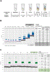
Materials and methods: Plasma samples from subjects with HPV+ OPSCC were collected. We developed a high-performance ddPCR assay designed to simultaneously target nine regions of the HPV16 genome.
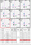
Results: The new assay termed 'ctDNA HPV16 Assessment using Multiple Probes' (CHAMP- 16) yielded significantly higher HPV16 counts compared to our previously validated 'Single-Probe' (SP) assay and a commercially available NavDx® assay. Analytical validation demonstrated that the CHAMP-16 assay had a limit of detection (LoD) of 4.1 copies per reaction, corresponding to < 1 genome equivalent (GE) of HPV16. When tested on plasma ctDNA from 21 patients with early-stage HPV+ OPSCC and known HPV16 ctDNA using the SP assay, all patients were positive for HPV16 ctDNA in both assays and the CHAMP-16 assay displayed 6.6-fold higher HPV16 signal on average. Finally, in a longitudinal analysis of samples from a patient with recurrent disease, the CHAMP-16 assay detected HPV16 ctDNA signal ∼ 20 months prior to the conventional SP assay.
Conclusion: Increased HPV16 signal detection using the CHAMP-16 assay suggests the potential for detection of recurrences significantly earlier than with conventional ddPCR assays in patients with HPV16+ OPSCC. Critically, this multi-probe approach maintains the cost-benefit advantage of ddPCR over next generation sequencing (NGS) approaches, supporting the cost-effectiveness of this assay for both large population screening and routine post-treatment surveillance.
Keywords: Anal cancer; Biomarker; Cell-free DNA; Cervical cancer; Circulating tumor DNA; HPV; Head and neck cancer; Liquid biopsy; Oropharyngeal cancer; ctDNA.
Published by Elsevier Ltd.
Conflict of interest statement
Declaration of Competing Interest The authors declare the following financial interests/personal relationships which may be considered as potential competing interests: Some of the authors are inventors of HPV ctDNA assay technology, for which the University of Michigan is pursuing patent protection.
Figures


Similar articles
-
HPV circulating tumoral DNA quantification by droplet-based digital PCR: A promising predictive and prognostic biomarker for HPV-associated oropharyngeal cancers.Int J Cancer. 2020 Aug 15;147(4):1222-1227. doi: 10.1002/ijc.32804. Epub 2019 Dec 18. Int J Cancer. 2020. PMID: 31756275
-
Detection of Circulating HPV16 DNA as a Biomarker for Cervical Cancer by a Bead-Based HPV Genotyping Assay.Microbiol Spectr. 2022 Apr 27;10(2):e0148021. doi: 10.1128/spectrum.01480-21. Epub 2022 Feb 28. Microbiol Spectr. 2022. PMID: 35225653 Free PMC article.
-
Analytical and clinical performance validation of HPV-SEQ, a novel NGS-based liquid biopsy platform for detection and quantification of human papilloma virus circulating tumor DNA.Oral Oncol. 2025 Aug;167:107445. doi: 10.1016/j.oraloncology.2025.107445. Epub 2025 Jun 26. Oral Oncol. 2025. PMID: 40578243
-
Circulating tumor DNA in human papillomavirus-associated oropharyngeal cancer management: A systematic review.Oral Oncol. 2025 May;164:107262. doi: 10.1016/j.oraloncology.2025.107262. Epub 2025 Mar 30. Oral Oncol. 2025. PMID: 40163959
-
Clinical outcomes of oropharyngeal squamous cell carcinoma stratified by human papillomavirus subtype: A systematic review and meta-analysis.Oral Oncol. 2024 Jan;148:106644. doi: 10.1016/j.oraloncology.2023.106644. Epub 2023 Nov 24. Oral Oncol. 2024. PMID: 38006690 Free PMC article.
Cited by
-
The Pivotal Role of Preclinical Animal Models in Anti-Cancer Drug Discovery and Personalized Cancer Therapy Strategies.Pharmaceuticals (Basel). 2024 Aug 9;17(8):1048. doi: 10.3390/ph17081048. Pharmaceuticals (Basel). 2024. PMID: 39204153 Free PMC article. Review.
-
Addressing knowledge gaps in HPV-related oropharyngeal cancer prevention: A call for action.Oral Oncol. 2025 Apr;163:107234. doi: 10.1016/j.oraloncology.2025.107234. Epub 2025 Mar 5. Oral Oncol. 2025. PMID: 40049067 Free PMC article. No abstract available.
-
Circulating Tumor HPV-DNA in the Management of HPV-Positive Oropharyngeal Carcinoma: A Systematic Review.Head Neck. 2025 Jun;47(6):1674-1679. doi: 10.1002/hed.28057. Epub 2025 Jan 23. Head Neck. 2025. PMID: 39846227 Free PMC article.
-
Prospective Phase II Trial of Definitive Chemoradiation and Concurrent Nivolumab in Locally Advanced p16+ Oropharynx Cancer.Adv Radiat Oncol. 2025 Jul 10;10(10):101832. doi: 10.1016/j.adro.2025.101832. eCollection 2025 Oct. Adv Radiat Oncol. 2025. PMID: 40837213 Free PMC article.
-
Toward a Next-Generation Liquid Biopsy for HPV+ HNSCC: Opportunities, Challenges, and the Road Ahead.Clin Cancer Res. 2025 Aug 14;31(16):3359-3361. doi: 10.1158/1078-0432.CCR-25-1488. Clin Cancer Res. 2025. PMID: 40552930
References
-
- Ramqvist T, Dalianis T. An epidemic of oropharyngeal squamous cell carcinoma (OSCC) due to human papillomavirus (HPV) infection and aspects of treatment and prevention. Anticancer Res 2011;31:1515–9. - PubMed
-
- Attner P, Du J, Näsman A, Hammarstedt L, Ramqvist T, Lindholm J, et al. The role of human papillomavirus in the increased incidence of base of tongue cancer. Int J Cancer 2010;126:2879–84. - PubMed
MeSH terms
Substances
Grants and funding
LinkOut - more resources
Full Text Sources
Medical

