. 2023 Jul:63:102765.
doi: 10.1016/j.redox.2023.102765. Epub 2023 May 28.

EETs alleviate alveolar epithelial cell senescence by inhibiting endoplasmic reticulum stress through the Trim25/Keap1/Nrf2 axis

Affiliations

EETs alleviate alveolar epithelial cell senescence by inhibiting endoplasmic reticulum stress through the Trim25/Keap1/Nrf2 axis

Chen-Yu Zhang et al. Redox Biol. 2023 Jul.

Abstract

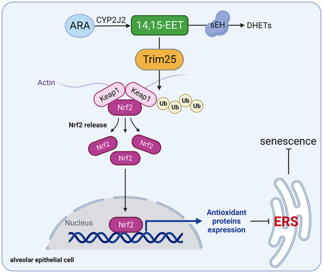
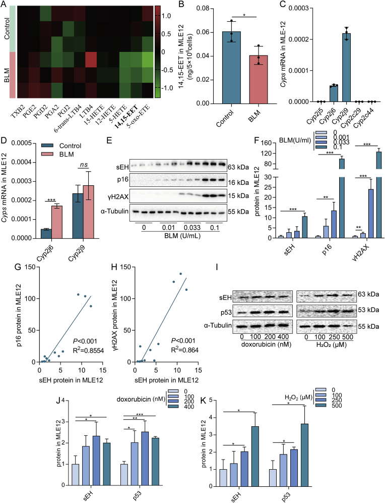
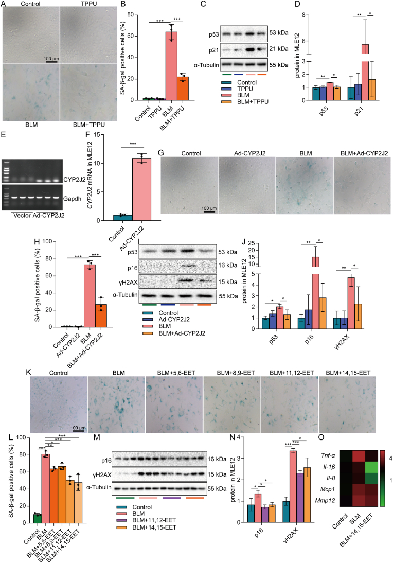
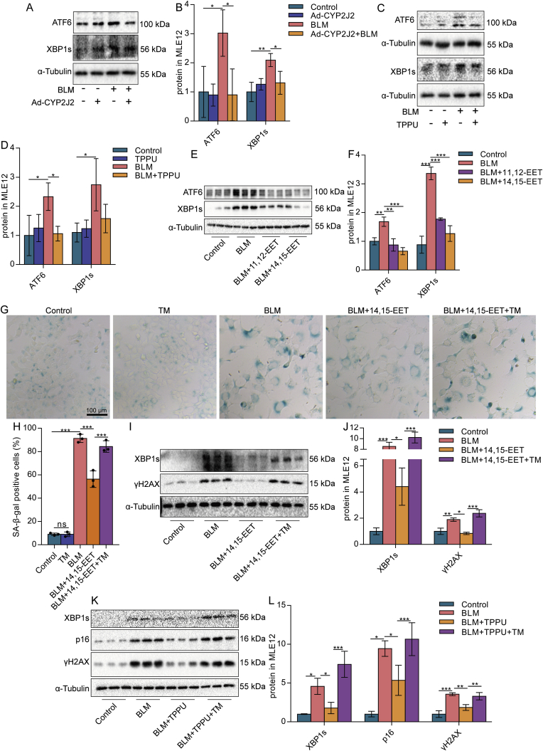
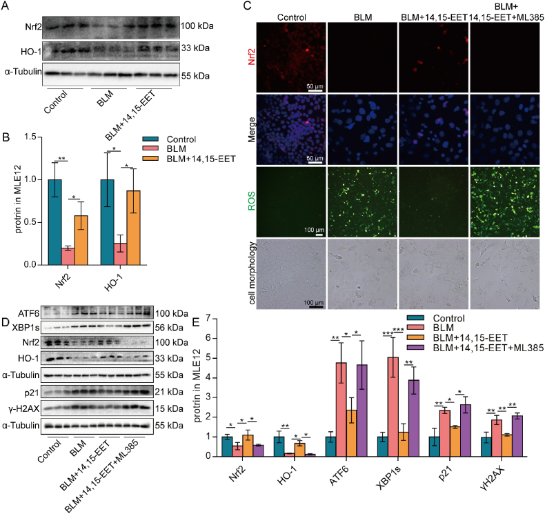
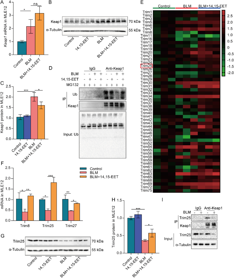
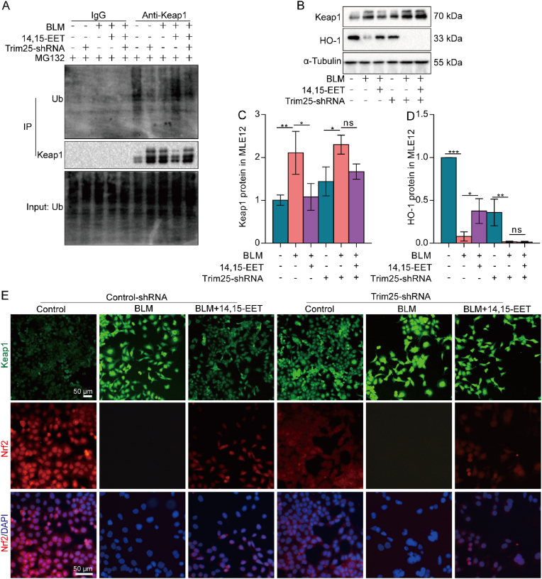
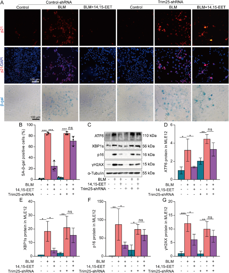
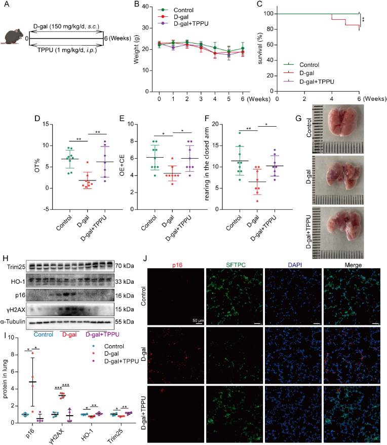
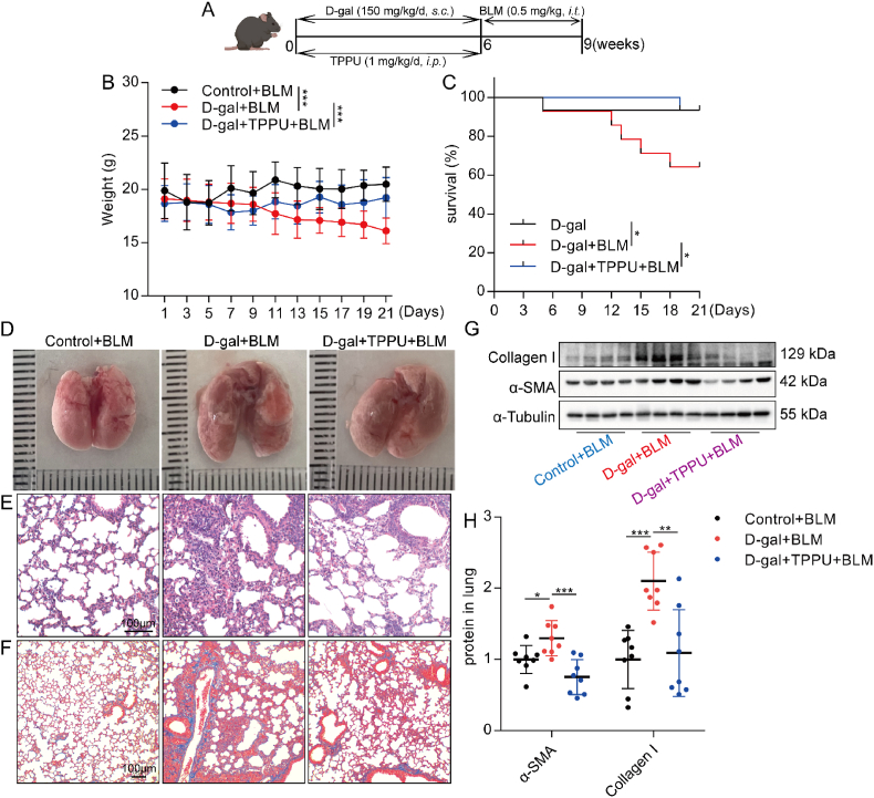
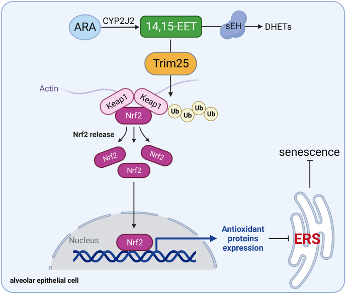
Alveolar epithelial cell (AEC) senescence is a key driver of a variety of chronic lung diseases. It remains a challenge how to alleviate AEC senescence and mitigate disease progression. Our study identified a critical role of epoxyeicosatrienoic acids (EETs), downstream metabolites of arachidonic acid (ARA) by cytochrome p450 (CYP), in alleviating AEC senescence. In vitro, we found that 14,15-EET content was significantly decreased in senescent AECs. Exogenous EETs supplementation, overexpression of CYP2J2, or inhibition of EETs degrading enzyme soluble epoxide hydrolase (sEH) to increase EETs alleviated AECs' senescence. Mechanistically, 14,15-EET promoted the expression of Trim25 to ubiquitinate and degrade Keap1 and promoted Nrf2 to enter the nucleus to exert an anti-oxidant effect, thereby inhibiting endoplasmic reticulum stress (ERS) and alleviating AEC senescence. Furthermore, in D-galactose (D-gal)-induced premature aging mouse model, inhibiting the degradation of EETs by Trifluoromethoxyphenyl propionylpiperidin urea (TPPU, an inhibitor of sEH) significantly inhibited the protein expression of p16, p21, and γH2AX. Meanwhile, TPPU reduced the degree of age-related pulmonary fibrosis in mice. Our study has confirmed that EETs are novel anti-senescence substances for AECs, providing new targets for the treatment of chronic lung diseases.

Keywords: Alveolar epithelial cells; Endoplasmic reticulum stress; Epoxyeicosatrienoic acids; Senescence; Tripartite motif-containing 25.


Conflict of interest statement

Declaration of competing interest The authors declare that they have no known competing financial interests or personal relationships that could have appeared to influence the work reported in this paper.

Figures

Image 1
Graphical abstract
Fig. 1
Fig. 1
Decreased EETs content is associated with the senescence of AECs. Heatmap showing the levels of arachidonic acid metabolites in MLE12 cells treated with BLM compared to normal saline. The content of metabolites was calculated in log2 (A, n = 3). Absolute quantification of 14,15-EET in MLE12 cells (B, n = 3). Cyps mRNA expression in MLE12 cells was detected by real-time PCR (C, n = 3). MLE12 cells were treated with BLM (0.1 U/mL) for 2 h. Cells were then washed with PBS and subsequently incubated for 22 h. The mRNA expression of Cyp2j6 and Cyp2j9 in MLE12 cells was detected by real-time PCR (D, n = 3). MLE12 cells were treated with different concentrations of BLM (0, 0.01, 0.033, and 0.1 U/mL) for 2 h. Cells were then washed with PBS and subsequently incubated for 46 h. The protein expression of sEH, p16, and γH2AX in MLE12 cells was detected by Western blot (E-F, n = 3). The linear associations between sEH and p16 or γH2AX protein expression were analyzed with Pearson correlations analysis (G–H). MLE12 cells were treated with different concentrations of doxorubicin (0, 100, 200, and 400 nM) or H2O2 (0, 100, 250, and 500 μM) for 2 h. Cells were then washed with PBS and subsequently incubated for 46 h. The protein expression of sEH and p53 in MLE12 cells was detected by Western blot (I–K, n = 3). Data are expressed as the mean ± SD. Differences among multiple groups were performed using ANOVA. Tukey's test was used as a post hoc test for pairwise comparisons. Comparisons between the two-group were made with an unpaired t-test. *P < 0.05, **P < 0.01, and ***P < 0.001.
Fig. 2
Fig. 2
Increasing EETs inhibits the senescence of AECs. MLE12 cells were treated with BLM (0.1 U/mL) for 2 h after TPPU (20 μM) intervention 1 h earlier, and then cultured for 46 h after BLM was removed. SA-β-gal staining was performed 48 h after BLM treatment (A-B, bar = 100 μm). The protein expression of p53 and p21 in MLE12 cells was detected by Western blot (C-D, n = 3). MLE12 cells were treated with BLM (0.1 U/mL) for 2 h after CYP2J2 overexpression, and then cultured for 46 h after BLM was removed. The expression of CYP2J2 mRNA in MLE12 cells 48 h after the adenovirus-CYP2J2 infection was detected by PCR (E-F, n = 3). SA-β-gal staining was performed 48 h after BLM treatment (G-H, bar = 100 μm). The protein expression of p53, p16, and γH2AX in MLE12 cells was detected by Western blot (I-J, n = 3). MLE12 cells were treated with BLM (0.1 U/mL) for 2 h after EETs (2.5 μM) intervention 10 min earlier, and then cultured for 46 h after BLM was removed. SA-β-gal staining was performed 48 h after BLM treatment (K-L, bar = 100 μm). The protein expression of p16 and γH2AX in MLE12 cells was detected by Western blot (M − N, n = 3). The mRNA expression Tnf-α, Il-1β, Il-8, Mcp1, and Mmp12 in MLE12 cells was detected by real-time PCR 24 h after BLM treatment (O, n = 3). Data are expressed as the mean ± SD. Differences among multiple groups were performed using ANOVA. Tukey's test was used as a post hoc test for pairwise comparisons. Comparisons between the two-group were made with an unpaired t-test. *P < 0.05, **P < 0.01, and ***P < 0.001.
Fig. 3
Fig. 3
EETs alleviate the senescence of AECs by reducing ERS. MLE12 cells were treated with BLM for 2 h after EETs intervention 10 min earlier, TPPU intervention 1 h earlier, or CYP2J2 overexpression, and then cultured for 46 h after BLM was removed. The protein expression of ATF6 and XBP1s in MLE12 cells was detected by Western blot (A-F, n = 3). MLE12 cells were treated with BLM for 2 h after 10 min of EETs intervention and 1 h after TPPU intervention, respectively. After culturing without BLM for 22 h, the cells were added to TM (0.25 μg/mL) for 24 h. SA-β-gal staining was performed 48 h after BLM treatment (G-H, bar = 100 μm). The protein expression of XBP1s and γH2AX in MLE12 cells was detected by Western blot (I-J, n = 3). The protein expression of XBP1s, p16, and γH2AX in MLE12 cells was detected by Western blot (K-L, n = 3). Data are expressed as the mean ± SD. Differences among multiple groups were performed using ANOVA. Tukey's test was used as a post hoc test for pairwise comparisons. *P < 0.05, **P < 0.01, and ***P < 0.001.
Fig. 4
Fig. 4
EETs alleviate ERS and senescence of AECs through Nrf2 anti-oxidant. MLE12 cells were treated with BLM for 2 h after EETs intervention 10 min earlier and cultured for 46 h after BLM was removed. The protein expression of Nrf2 and HO-1 in MLE12 cells was detected by Western blot (A-B, n = 3). The fluorescence intensity of Nrf2 was detected by immunofluorescence (C, bar = 50 μm). The ROS in MLE12 cells were detected by a ROS kit (C, bar = 100 μm). Cell morphology is photographed through a microscope (C, bar = 100 μm). Cells were treated with ML385 (5 μM) for 1 h before the treatment with BLM. ATF6, XBP1s, Nrf2, HO-1, p21, and γH2AX protein expression in MLE12 cells was detected by Western blot (D-E, n = 3). Data are expressed as the mean ± SD. Differences among multiple groups were performed using ANOVA. Tukey's test was used as a post hoc test for pairwise comparisons. *P < 0.05, **P < 0.01, and ***P < 0.001.
Fig. 5
Fig. 5
EETs increase Keap1 ubiquitination level and E3 ubiquitin ligase Trim25 expression.Keap1 mRNA expression in MLE12 cells was detected by real-time PCR 24 h after BLM treatment (A, n = 3). Keap1 protein expression was detected by Western blot 48 h after BLM treatment (B–C, n = 3). Keap1 ubiquitination level was detected by Co-IP in MLE12 cells (D). Trims mRNA expression in MLE12 cells was detected by real-time PCR 24 h after BLM treatment (E, n = 3). The mRNA expression of Trim8, Trim25, and Trim27 was detected by real-time PCR (F, n = 3). Trim25 protein expression was detected by Western blot 48 h after BLM treatment (G-H, n = 3). The interaction between Keap1 and Trim25 was detected by Co-IP in MLE12 cells (I). Data are expressed as the mean ± SD. Differences among multiple groups were performed using ANOVA. Tukey's test was used as a post hoc test for pairwise comparisons. *P < 0.05, **P < 0.01, and ***P < 0.001.
Fig. 6
Fig. 6
EETs promote Nrf2 into the nucleus by ubiquitination of Keap1 by Trim25. Lentivirus was used to silence Trim25 in MLE12 cells. Keap1 ubiquitination level was detected by Co-IP in MLE12 cells (A). The protein expression of Keap1 and HO-1 in MLE12 cells was detected by Western blot (B-D, n = 3). The fluorescence intensity of Keap1 and Nrf2 was detected by immunofluorescence (E, bar = 50 μm). Data are expressed as the mean ± SD. Differences among multiple groups were performed using ANOVA. Tukey's test was used as a post hoc test for pairwise comparisons. *P < 0.05, **P < 0.01, and ***P < 0.001.
Fig. 7
Fig. 7
EETs alleviate AEC senescence by inhibiting ERS through Trim25/Keap1/Nrf2. Lentivirus was used to silence Trim25 in MLE12 cells. The fluorescence intensity of p21 was detected by immunofluorescence (A, bar = 100 μm). SA-β-gal staining was performed 48 h after BLM treatment (A-B, bar = 100 μm). The protein expression of ATF6, XBP1s, p16, and γH2AX in MLE12 cells was detected by Western blot (C-G, n = 3). Data are expressed as the mean ± SD. Differences among multiple groups were performed using ANOVA. Tukey's test was used as a post hoc test for pairwise comparisons. *P < 0.05, **P < 0.01, and ***P < 0.001.
Fig. 8
Fig. 8
Inhibition of EETs degradation alleviates D-gal-induced lung senescence in aging mice. C57BL/6 J mice were given a daily subcutaneous injection of D-gal (150 mg/kg) in the neck and back to establish the aging model and a daily intraperitoneal injection of TPPU (1 mg/kg) (A). Weight change of mice (B, n = 28). The survival rate was expressed as Kaplan Meier survival curves (C, n = 28). Elevated plus-maze test, OT% (Open arm retention time ratio), OE + CE (The total number of times the open and closed arms were entered), and rearing (D–F). The appearance of lung tissue (G). The protein expression of Trim 25, HO-1, p16, and γH2AX in the lung was detected by Western blot (H–I, n = 7). The fluorescence intensity of p16 and SFTPC was detected by immunofluorescence (J, bar = 50 μm). Data are expressed as the mean ± SD. Differences among multiple groups were performed using ANOVA. Tukey's test was used as a post hoc test for pairwise comparisons. Survival data were analyzed using the log-rank test. *P < 0.05, **P < 0.01, and ***P < 0.001.
Fig. 9
Fig. 9
Inhibition of EETs degradation alleviates experimental pulmonary fibrosis in aging mice. D-gal-induced aging mice were given a tracheal injection of BLM (0.5 mg/kg) (A). Weight change of mice (B, n = 14–15). The survival rate was expressed as Kaplan Meier survival curves (C, n = 14–15). The appearance of lung tissue (D). HE staining of lung tissue (E, bar = 100 μm). Masson staining of lung tissue (F, bar = 100 μm). The protein expression of Collagen I and α-SMA in the lung was detected by Western blot (I-J, n = 8). Data are expressed as the mean ± SD. Differences among multiple groups were performed using ANOVA. Tukey's test was used as a post hoc test for pairwise comparisons. Survival data were analyzed using the log-rank test. *P < 0.05, **P < 0.01, and ***P < 0.001.
Fig. 10
Fig. 10
The schematic diagram for EETs alleviate AEC senescence. 14,15-EET degrades Keap1 through Trim25 ubiquitination and promotes Nrf2 nuclear translocation to play an anti-oxidant role, inhibits ERS, and attenuates AEC senescence.

Similar articles

Cited by

References

    1. Dzau V.J., Inouye S.K., Rowe J.W., Finkelman E., Yamada T. Enabling healthful aging for all - the national Academy of medicine grand challenge in healthy longevity. N. Engl. J. Med. 2019;381:1699–1701. doi: 10.1056/NEJMp1912298. - DOI - PubMed
    1. Schneider J.L., Rowe J.H., Garcia-de-Alba C., Kim C.F., Sharpe A.H., Haigis M.C. The aging lung: physiology, disease, and immunity. Cell. 2021;184:1990–2019. doi: 10.1016/j.cell.2021.03.005. - DOI - PMC - PubMed
    1. Calcinotto A., Kohli J., Zagato E., Pellegrini L., Demaria M., Alimonti A. Cellular senescence: aging, cancer, and injury. Physiol. Rev. 2019;99:1047–1078. doi: 10.1152/physrev.00020.2018. - DOI - PubMed
    1. Di Micco R., Krizhanovsky V., Baker D., d'Adda di Fagagna F. Cellular senescence in ageing: from mechanisms to therapeutic opportunities. Nat. Rev. Mol. Cell Biol. 2021;22:75–95. doi: 10.1038/s41580-020-00314-w. - DOI - PMC - PubMed
    1. Childs B.G., Baker D.J., Wijshake T., Conover C.A., Campisi J., van Deursen J.M. Senescent intimal foam cells are deleterious at all stages of atherosclerosis. Science. 2016;354:472–477. doi: 10.1126/science.aaf6659. - DOI - PMC - PubMed

Publication types

MeSH terms

LinkOut - more resources