. 2023 Aug:314:104087.
doi: 10.1016/j.resp.2023.104087. Epub 2023 Jun 2.

Arginine vasopressin potentiates inspiratory bursting in hypoglossal motoneurons of neonatal mice

Affiliations

Arginine vasopressin potentiates inspiratory bursting in hypoglossal motoneurons of neonatal mice

K N Bolte et al. Respir Physiol Neurobiol. 2023 Aug.

Abstract

Vasopressin (AVP) acts as a neurotransmitter and its activity can potentiate respiratory activity. Hypoglossal (XII) motoneurons that innervate the tongue express V1a vasopressin receptors, which are excitatory. Therefore, we hypothesized that V1a receptor activation at XII motoneurons would potentiate inspiratory bursting. We developed this study to determine whether AVP can potentiate inspiratory bursting in rhythmic medullary slice preparations in neonatal (postnatal, P0-5) mice. Bath or local application of AVP potentiated inspiratory bursting compared to baseline XII inspiratory burst amplitude. Antagonizing V1a receptors revealed significant attenuation of the AVP-mediated potentiation of inspiratory bursting, while antagonism of oxytocin receptors (at which AVP has similar binding affinity) revealed a trend to attenuate AVP-mediated potentiation of inspiratory bursting. Finally, we discovered that the AVP-mediated potentiation of inspiratory bursting increases significantly with postnatal maturation from P0-5. Overall, these data support that AVP potentiates inspiratory bursting directly at XII motoneurons.

Keywords: Anti-diuretic hormone; Breathing; Hypoglossal; Inspiratory pattern; Motor neurons; Neuromodulation; Oxytocin; Respiration; V1a.


Conflict of interest statement

Declaration of Competing Interest The authors declare no competing interests.

Figures
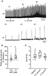
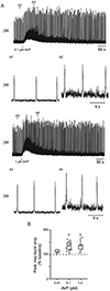
Figure 1.
Figure 1.. Effects of bath application of AVP on inspiratory bursting in the rhythmic slice preparation.
A) Rectified and integrated recording of XII nerve activity demonstrating inspiratory burst parameter changes during bath application of AVP at 0.01 and 0.1 μM (arrowhead denotes addition of AVP to the bathing solution). Expanded time scale of inspiratory bursts during baseline (A1), 0.01 μM AVP (A2) and 0.1 μM AVP (A3) taken at the times denoted by the arrowheads on the continuous trace. Group data (box and whisker plot: interquartile range and median, 9th and 91st confidence intervals) and individual data points (open circles) for burst amplitude (B) and for burst period (C) averaged for five minutes from XII nerve rootlet recordings where AVP was applied in the bath for 10 minutes prior to measurements, n = 11 (baseline and 0.01 μM AVP) and 10 (0.1 μM AVP). *, p < 0.05, mixed-effects analysis with Tukey’s multiple comparisons post-hoc test.
Figure 2.
Figure 2.. Effects of local application of AVP on inspiratory bursting in the rhythmic slice preparation.
A) Rectified and integrated recording of XII nerve activity demonstrating inspiratory burst parameter changes during 15 sec local application of AVP at 0.1 (top trace) and 1 μM (bottom trace), black bar denotes AVP application. Expanded time scale of inspiratory bursts during baseline (A1), 0.1 μM AVP (A2), baseline (A3), and 1 μM AVP (A4) denoted by the arrowheads on the continuous trace. B) Group data (box and whisker plot: interquartile range and median, 9th and 91st confidence intervals) and individual data points (open circles), n = 6 (0.01 μM AVP), 13 (0.1 μM AVP), and 5 (1 μM AVP). *, p < 0.05, mixed-effects analysis with Tukey’s multiple comparisons post-hoc test.
Figure 3.
Figure 3.. Effects of local application of AVP V1a receptor antagonist (d(CH2)51,Tyr(Me)2,Arg8)-Vasopressin on AVP-mediated potentiation of inspiratory bursting in the rhythmic slice preparation.
A) Rectified and integrated recording of XII nerve activity demonstrating inspiratory burst parameter changes during 15 sec local application of 1 μM AVP (top trace), followed by a 60 s local pre-application of (d(CH2)51,Tyr(Me)2,Arg8)-Vasopressin (V1a receptor antagonist) prior to a 15 sec local application of 1 μM AVP (middle trace), and then a third 15 sec local application of 1 μM AVP (bottom trace), black bar denotes AVP application, red bar denotes (d(CH2)51,Tyr(Me)2,Arg8)-Vasopressin application. Expanded time scale of inspiratory bursts during baseline (A1), 1 μM AVP (A2), baseline (A3), and 1 μM AVP after (d(CH2)51,Tyr(Me)2,Arg8)-Vasopressin (A4), baseline (A5), 1 μM AVP (A6) denoted by the arrowheads on the continuous trace. B) Group data (box and whisker plot: interquartile range and median, 9th and 91st confidence intervals) and individual data points each experiment linked by lines between open circles, n = 8 (first 1 μM AVP) and 10 (antagonist pre-application and washout 1 μM AVP). *, p < 0.05, mixed-effects analysis with Tukey’s multiple comparisons post-hoc test.
Figure 4.
Figure 4.. Effects of local application of oxytocin receptor antagonist L-371,257 on AVP-mediated potentiation of inspiratory bursting in the rhythmic slice preparation.
A) Rectified and integrated recording of XII nerve activity demonstrating inspiratory burst parameter changes during 15 sec local application of 1 μM AVP (top trace), followed by a 60 s local pre-application of L-371,257 (oxytocin receptor antagonist) prior to a 15 sec local application of 1 μM AVP (middle trace), and then a third 15 sec local application of 1 μM AVP (bottom trace), black bar denotes AVP application, red bar denotes L-371,257 application. Expanded time scale of inspiratory bursts during baseline (A1), 1 μM AVP (A2), baseline (A3), and 1 μM AVP after L-371,257 (A4), baseline (A5), 1 μM AVP (A6) denoted by the arrowheads on the continuous trace. B) Group data (box and whisker plot: interquartile range and median, 9th and 91st confidence intervals) and individual data points each experiment linked by lines between open circles, n = 7. P > 0.05, mixed-effects analysis.
Figure 5.
Figure 5.. Effects of local application of AVP on inspiratory bursting in the rhythmic slice preparation compared to male or female sex.
A) Rectified and integrated recording of XII nerve activity demonstrating inspiratory burst parameter changes during 15 sec local application of 1 μM AVP in female P2 (top trace) and male P2 (bottom trace) rhythmic medullary slice preparations. Expanded time scale of inspiratory bursts during baseline (A1), 1 μM AVP (A2) in a female preparation, baseline (A3), and 1 μM AVP (A4) in a male preparation, denoted by the arrowheads on the continuous trace. B) Group data (box and whisker plot: interquartile range and median, 9th and 91st confidence intervals) and individual data points (open circles). N = 18 (female) and 18 (male). P > 0.05 (paired t-test).
Figure 6.
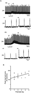
Figure 6.. Effects of local application of AVP on inspiratory bursting in the rhythmic slice preparation compared to postnatal age.
A) Rectified and integrated recording of XII nerve activity demonstrating inspiratory burst parameter changes during 15 sec local application of 1 μM AVP in P0 (top trace) and P5 (bottom trace) rhythmic medullary slice preparations. Expanded time scale of inspiratory bursts during baseline (A1), 1 μM AVP (A2) in a P0 preparation, and baseline (A3), and 1 μM AVP (A4) in a P5 preparation, denoted by the arrowheads on the continuous trace. B) Group data showing the potentiation of XII inspiratory peak burst amplitude mediated by AVP (1 μM) in rhythmic medullary slice preparations as a function of postnatal age between P0-5 (n = 36).

Similar articles

References

    1. Adachi T, Huxtable AG, Fang X, and Funk GD. Substance P modulation of hypoglossal motoneuron excitability during development: changing balance between conductances. J Neurophysiol 104: 854–872, 2010. - PubMed
    1. Aulino EA, and Caldwell HK. Subtle sex differences in vasopressin mRNA expression in the embryonic mouse brain. J Neuroendocrinol 32: e12835, 2020. - PMC - PubMed
    1. Bankir L, Bichet DG, and Morgenthaler NG. Vasopressin: physiology, assessment and osmosensation. J Intern Med 282: 284–297, 2017. - PubMed
    1. Barberis C, Mouillac B, and Durroux T. Structural bases of vasopressin/oxytocin receptor function. J Endocrinol 156: 223–229, 1998. - PubMed
    1. Bayliss DA, Viana F, and Berger AJ. Mechanisms underlying excitatory effects of thyrotropin-releasing hormone on rat hypoglossal motoneurons in vitro. J Neurophysiol 68: 1733–1745, 1992. - PubMed

Publication types

LinkOut - more resources