. 2023 Sep;23(9):1411-1424.
doi: 10.1016/j.ajt.2023.05.025. Epub 2023 Jun 1.

The role of interleukin-21 in COVID-19 vaccine-induced B cell-mediated immune responses in patients with kidney disease and kidney transplant recipients

Collaborators, Affiliations

The role of interleukin-21 in COVID-19 vaccine-induced B cell-mediated immune responses in patients with kidney disease and kidney transplant recipients

S Reshwan K Malahe et al. Am J Transplant. 2023 Sep.

Abstract

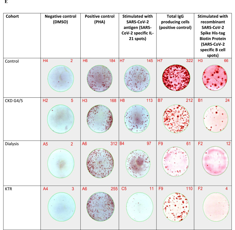
T-cell-mediated help to B cells is required for the development of humoral responses, in which the cytokine interleukin (IL)-21 is key. Here, we studied the mRNA-1273 vaccine-induced SARS-CoV-2-specific memory T-cell IL-21 response, memory B cell response, and immunoglobulin (Ig)G antibody levels in peripheral blood at 28 days after the second vaccination by ELISpot and the fluorescent bead-based multiplex immunoassay, respectively. We included 40 patients with chronic kidney disease (CKD), 34 patients on dialysis, 63 kidney transplant recipients (KTR), and 47 controls. We found that KTR, but not patients with CKD and those receiving dialysis, showed a significantly lower number of SARS-CoV-2-specific IL-21 producing T cells than controls (P < .001). KTR and patients with CKD showed lower numbers of SARS-CoV-2-specific IgG-producing memory B cells when compared with controls (P < .001 and P = .01, respectively). The T-cell IL-21 response was positively associated with the SARS-CoV-2-specific B cell response and the SARS-CoV-2 spike S1-specific IgG antibody levels (both Pearson r = 0.5; P < .001). In addition, SARS-CoV-2-specific B cell responses were shown to be IL-21 dependent. Taken together, we show that IL-21 signaling is important in eliciting robust B cell-mediated immune responses in patients with kidney disease and KTR.

Keywords: COVID-19; SARS-CoV-2; immune responses; interleukin-21; kidney transplant recipients; patients with kidney disease; vaccination.


Conflict of interest statement

Declaration of competing interest The authors declare that they have no known competing financial interests or personal relationships that could have appeared to influence the work reported in this paper.

Figures

Figure 1
Figure 1
SARS-CoV-2–specific memory T-cell IL-21 and memory B cell response measured by ELISpot. Data are presented in box-and-whisker plots. The horizontal line and numbers within the whisker indicate the medians and the tops and bottoms indicate interquartile ranges. Mann-Whitney U tests were applied to compare medians. LLoD stands for the lower limit of detection and was 3.3 spots (or 0.52 if log10-transformed) for memory T-cell IL-21 response and 5 spots (or 0.7 if log10-transformed) for memory B cell response. Each symbol represents an individual. SARS-CoV-2–specific memory T-cell IL-21 response at (A) baseline and (B) 28 days after the second vaccination. SARS-CoV-2–specific memory B cell response at (C) baseline (n = subset of participants) and (D) 28 days after the second vaccination (n = subset of participants). (E) Illustration of spots measured by ELISpot. The top right number in red indicates the number of spots measured by the ELISpot assay (IL-21 producing cells per 300 000 PBMCs/well, total IgG–producing cells per 10 000 PBMCs/well or SARS-CoV-2–specific B cell spots per 200 000 PBMCs/well) and the top left number in red indicates the location on the 96-well PVDF plate. CKD, chronic kidney disease; KTR, kidney transplant recipient; PBMC, peripheral blood mononuclear cell.
Figure 1
Figure 1
SARS-CoV-2–specific memory T-cell IL-21 and memory B cell response measured by ELISpot. Data are presented in box-and-whisker plots. The horizontal line and numbers within the whisker indicate the medians and the tops and bottoms indicate interquartile ranges. Mann-Whitney U tests were applied to compare medians. LLoD stands for the lower limit of detection and was 3.3 spots (or 0.52 if log10-transformed) for memory T-cell IL-21 response and 5 spots (or 0.7 if log10-transformed) for memory B cell response. Each symbol represents an individual. SARS-CoV-2–specific memory T-cell IL-21 response at (A) baseline and (B) 28 days after the second vaccination. SARS-CoV-2–specific memory B cell response at (C) baseline (n = subset of participants) and (D) 28 days after the second vaccination (n = subset of participants). (E) Illustration of spots measured by ELISpot. The top right number in red indicates the number of spots measured by the ELISpot assay (IL-21 producing cells per 300 000 PBMCs/well, total IgG–producing cells per 10 000 PBMCs/well or SARS-CoV-2–specific B cell spots per 200 000 PBMCs/well) and the top left number in red indicates the location on the 96-well PVDF plate. CKD, chronic kidney disease; KTR, kidney transplant recipient; PBMC, peripheral blood mononuclear cell.
Figure 2
Figure 2
Correlation between tacrolimus trough level within 1 month before baseline and vaccine–induced memory T-cell IL-21 response in kidney transplant recipients (KTRs) at 28 days after the second vaccination (Pearson r = −0.4; P =.02). The horizontal dotted line represents the lower limit of detection of memory T-cell IL-21 response (= 3.3 spots or 0.52 if log10-transformed).
Figure 3
Figure 3
Correlation between vaccine–induced memory T-cell IL-21 response and memory B cell response at 28 days after the second vaccination (Pearson r = 0.5; P <.001). The horizontal dotted line represents the lower limit of detection of memory B cell response (=5 spots or 0.7 if log10-transformed) and the vertical dotted line represents the lower limit of detection of memory T-cell IL-21 response (=3.3 spots or 0.52 if log10-transformed).
Figure 4
Figure 4
Correlation between vaccine–induced memory T-cell IL-21 response and antibody responses. (A) Correlation between memory T-cell IL-21 response and S1 IgG antibody levels both at 28 days after the second vaccination (Pearson r = 0.5). (B) Correlation between memory T-cell IL-21 response at 28 days after the second vaccination and S1 IgG antibody levels at 6 months after the second vaccination (Pearson r = 0.5). The horizontal dotted line represents the cutoff value for being a serological responder (≥10 BAU/mL or ≥1 if log10-transformed), and the vertical dotted line represents the lower limit of detection of IL-21 response (=3.3 spots or 0.52 if log10-transformed). Each symbol represents an individual.
Figure 5
Figure 5
Correlation between vaccine–induced memory B cell response and antibody responses. (A) Correlation between memory B cell response and S1 IgG antibody levels at 28 days after the second vaccination (Pearson r = 0.5). (B) Correlation between memory B cell response at 28 days after the second vaccination and S1 IgG antibody levels at 6 months after the second vaccination (Pearson r = 0.4). The horizontal dotted line represents the cutoff value for being a serological responder (≥10 BAU/mL or ≥1 if log10-transformed), and the vertical dotted line represents the lower limit of detection of memory B cell response (=5 spots or 0.7 if log10-transformed). Each symbol represents an individual.
Figure 6
Figure 6
Effect of blocking IL-21 by adding an IL-21 receptor antagonist (ATR-107) on the vaccine–induced SARS-CoV-2–specific memory B cell response. (A) PBMCs from 4 health care workers were used and treated under the same condition of the B cell ELISpot assay and measured in the IL-21 ELISpot assay, in which PBMCs were stimulated with the standard concentration of IL-2+R848 to demonstrate that IL-2+R848 was able to induce the secretion of IL-21. The IL-21R antagonist was added in 4 different concentrations (1, 5, 10, and 20 μg/mL) to the B cell ELISpot in samples from 7 health care workers and 5 controls subjects of the RECOVAC Immune Response study, who were vaccinated twice with a COVID-19 mRNA vaccine. (B) SARS-CoV-2–specific memory B cell response at 28 days after the second vaccination. (C) Total IgG–producing cells at 28 days after the second vaccination, which served as the positive control for the B cell ELISpot. In some experiments, the number of participants was <12 owing to a lack of PBMCs or technical failure. Data are presented in box-and-whisker plots. The horizontal line and numbers within the whisker indicate the medians, and the tops and bottoms indicate interquartile ranges. Mann-Whitney U tests were applied to compare medians. LLoD stands for the lower limit of detection and was 5 spots (or 0.7 if log10-transformed) for memory B cell response. Each symbol represents an individual.

References

    1. Khoury D.S., Cromer D., Reynaldi A., et al. Neutralizing antibody levels are highly predictive of immune protection from symptomatic SARS-CoV-2 infection. Nat Med. 2021;27(7):1205–1211. doi: 10.1038/s41591-021-01377-8. - DOI - PubMed
    1. Kho M.M.L., Reinders M.E.J., Baan C.C., et al. The RECOVAC IR study: the immune response and safety of the mRNA-1273 COVID-19 vaccine in patients with chronic kidney disease, on dialysis or living with a kidney transplant. Nephrol Dial Transplant. 2021;36(9):1761–1764. doi: 10.1093/ndt/gfab186. - DOI - PMC - PubMed
    1. Sanders J.F., Messchendorp A.L., de Vries R.D., et al. Antibody and T-cell responses 6 months after COVID-19 mRNA-1273 vaccination in patients with chronic kidney disease, on dialysis, or living with a kidney transplant. Clin Infect Dis. 2023;76(3):e188–e199. doi: 10.1093/cid/ciac557. - DOI - PMC - PubMed
    1. Leonard W.J., Wan C.K. IL-21 signaling in immunity. F1000Res. 2016;5 doi: 10.12688/f1000research.7634.1. F1000 FacultyRev-224. - DOI - PMC - PubMed
    1. Ostiguy V., Allard E.L., Marquis M., Leignadier J., Labrecque N. IL-21 promotes T lymphocyte survival by activating the phosphatidylinositol-3 kinase signaling cascade. J Leukoc Biol. 2007;82(3):645–656. doi: 10.1189/jlb.0806494. - DOI - PubMed

Publication types