. 2023 Jun 3;15(1):103.
doi: 10.1186/s13195-023-01251-4.

APOE4 carrier status determines association between white matter disease and grey matter atrophy in early-stage dementia

Affiliations

APOE4 carrier status determines association between white matter disease and grey matter atrophy in early-stage dementia

Ashwati Vipin et al. Alzheimers Res Ther. .

Abstract

Background: White matter hyperintensities, a neuroimaging marker of small-vessel cerebrovascular disease and apolipoprotein ε4 (APOE4) allele, are important dementia risk factors. However, APOE4 as a key effect modifier in the relationship between white matter hyperintensities and grey matter volume needs further exploration.

Methods: One hundred ninety-two early-stage dementia (including mild cognitive impairment and mild dementia) and 259 cognitively unimpaired participants from a neurocognitive research cohort with neuroimaging data, APOE genotyping, and neuropsychological assessments were studied. We investigated independent and interactive effects of white matter hyperintensities and APOE4 on whole-brain voxel-wise grey matter volume using voxel-based morphometry (uncorrected p < 0.001; minimum cluster size = 100 voxels). We further assessed interactive effects between APOE4 and white matter hyperintensities on global cognition, memory, and executive function in early-stage dementia and cognitively unimpaired participants.

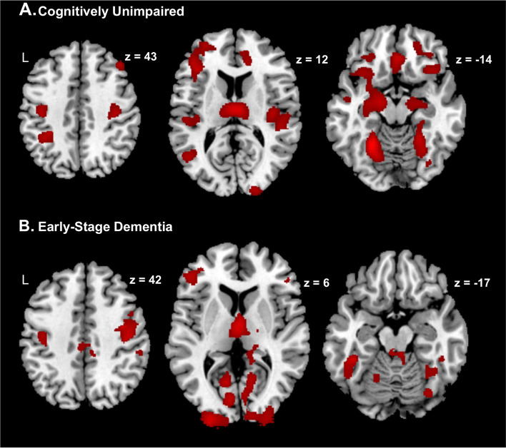
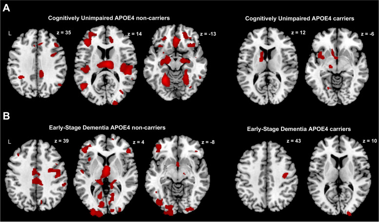
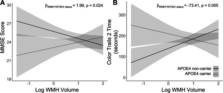
Results: Independent of APOE4 status, higher white matter hyperintensity load was associated with greater grey matter atrophy across frontal, parietal, temporal, and occipital lobes in cognitively unimpaired and early-stage dementia subjects. However, interaction analyses and independent sample analyses revealed that APOE4 non-carriers demonstrated greater white matter hyperintensity-associated grey matter atrophy compared to APOE4 carriers in both cognitively unimpaired and early-stage dementia groups. Additional confirmatory analyses among APOE4 non-carriers demonstrated that white matter hyperintensities resulted in widespread grey matter loss. Analyses of cognitive function demonstrated that higher white matter hyperintensity load was associated with worse global (Mini-Mental State Examination, Montreal Cognitive Assessment) and executive function (Color Trails 2) in APOE4 non-carriers compared to APOE4 carriers in early-stage dementia but not cognitively unimpaired participants.

Conclusions: The association between white matter hyperintensities and grey matter loss is more pronounced in APOE4 non-carriers than APOE4 carriers in the cognitively unimpaired and early-stage dementia stages. Furthermore, white matter hyperintensity presence results in poorer executive function in APOE4 non-carriers compared to APOE4 carriers. This finding may have significant impact on the design of clinical trials with disease modifying therapies.

Keywords: APOE4; Cognition; Cognitively normal; Dementia; Grey matter; White matter hyperintensity.


Conflict of interest statement

The authors declare no competing interests.

Figures

Fig. 1
Fig. 1
Negative influence of white matter hyperintensity load on voxel-wise grey matter volume in cognitively unimpaired and early-stage dementia participants. In both cognitively unimpaired (A) and early dementia participants (B), increasing WMH load was associated with lower voxel-wise GMV across frontal, parietal, temporal, and occipital regions. Clusters showing GMV loss related to WMH are shown in red. There were no positive associations between WMH load and voxel-wise GMV. Results are shown at the uncorrected p < 0.001 height threshold with an extent threshold of 100 voxels. Results are displayed on representative sections of the MNI template brain. WMH, white matter hyperintensity; GMV, grey matter volume
Fig. 2
Fig. 2
Among cognitively unimpaired and early-stage dementia participants, APOE4 carrier status influences the effect of white matter hyperintensity load on voxel-wise grey matter volume. In both cognitively unimpaired (A) and early-stage dementia participants (B) APOE4 non-carriers, increasing WMH load was associated with widespread lower voxel-wise GMV across frontal, parietal, temporal, and occipital regions only in APOE4 non-carriers. On the other hand, in cognitively unimpaired (A) APOE4 carriers, increasing WMH load was associated with lower voxel-wise GMV only in the left middle and superior temporal pole, caudate and lingual gyrus and in early-stage dementia participants (B) only in right precentral and superior occipital gyrus. Clusters showing GMV loss related to WMH are shown in red. Results are shown at the uncorrected p < 0.001 height threshold with an extent threshold of 100 voxels. Results are displayed on representative sections of the MNI template brain. WMH, white matter hyperintensity; GMV, grey matter volume; APOE4, apolipoprotein E4
Fig. 3
Fig. 3
White matter hyperintensity load among early-stage dementia APOE4 non-carriers relates to worse cognition than APOE4 carriers. In early-stage dementia participants, there was a significant interaction effect between WMH load and APOE4 status on cognition. Increasing white matter hyperintensity load was associated with (A) lower MMSE scores and (B) poorer Color Trails 2 performance in APOE4 non-carriers compared to APOE4 carriers. The solid line refers to APOE4 non-carriers while the dashed line refers to APOE4 carriers. APOE4, apolipoprotein E4; MMSE, mini-mental state examination; WMH, white matter hyperintensity

References

    1. Tangalos EG, Petersen RC. Mild cognitive impairment in geriatrics. Clin Geriatr Med. 2018;34(4):563–589. doi: 10.1016/j.cger.2018.06.005. - DOI - PubMed
    1. Li L, Wang Y, Yan J, Chen Y, Zhou R, Yi X, et al. Clinical predictors of cognitive decline in patients with mild cognitive impairment: the Chongqing aging study. J Neurol. 2012;259(7):1303–1311. doi: 10.1007/s00415-011-6342-0. - DOI - PubMed
    1. Campbell NL, Unverzagt F, LaMantia MA, Khan BA, Boustani MA. Risk factors for the progression of mild cognitive impairment to dementia. Clin Geriatr Med. 2013;29(4):873. doi: 10.1016/j.cger.2013.07.009. - DOI - PMC - PubMed
    1. Ramirez J, McNeely AA, Berezuk C, Gao F, Black SE. Dynamic progression of white matter hyperintensities in Alzheimer’s disease and normal aging: results from the Sunnybrook dementia study. Front Aging Neurosci. 2016;8:62. doi: 10.3389/fnagi.2016.00062. - DOI - PMC - PubMed
    1. Tosto G, Zimmerman ME, Carmichael OT, Brickman AM. Predicting aggressive decline in mild cognitive impairment: the importance of white matter hyperintensities. JAMA Neurol. 2014;71(7):872–877. doi: 10.1001/jamaneurol.2014.667. - DOI - PMC - PubMed

Publication types

MeSH terms