. 2023 Jun 3;22(1):92.
doi: 10.1186/s12943-023-01789-9.

CXCR2 expression during melanoma tumorigenesis controls transcriptional programs that facilitate tumor growth

Affiliations

CXCR2 expression during melanoma tumorigenesis controls transcriptional programs that facilitate tumor growth

J Yang et al. Mol Cancer. .

Abstract

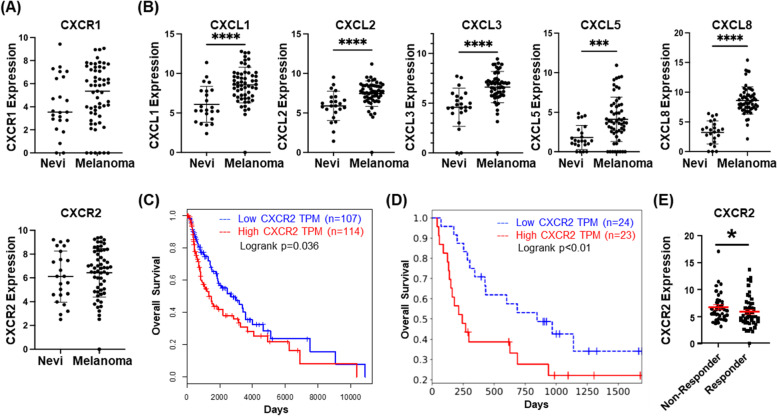
Background: Though the CXCR2 chemokine receptor is known to play a key role in cancer growth and response to therapy, a direct link between expression of CXCR2 in tumor progenitor cells during induction of tumorigenesis has not been established.

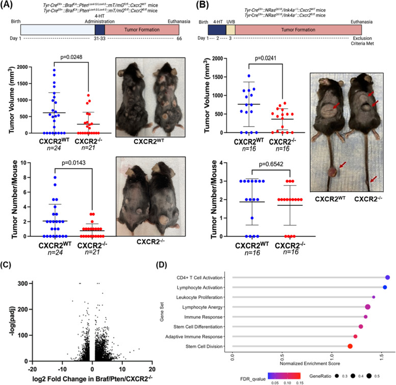
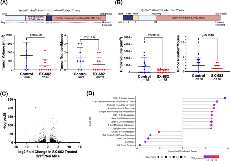
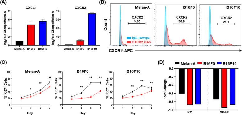
Methods: To characterize the role of CXCR2 during melanoma tumorigenesis, we generated tamoxifen-inducible tyrosinase-promoter driven BrafV600E/Pten-/-/Cxcr2-/- and NRasQ61R/INK4a-/-/Cxcr2-/- melanoma models. In addition, the effects of a CXCR1/CXCR2 antagonist, SX-682, on melanoma tumorigenesis were evaluated in BrafV600E/Pten-/- and NRasQ61R/INK4a-/- mice and in melanoma cell lines. Potential mechanisms by which Cxcr2 affects melanoma tumorigenesis in these murine models were explored using RNAseq, mMCP-counter, ChIPseq, and qRT-PCR; flow cytometry, and reverse phosphoprotein analysis (RPPA).

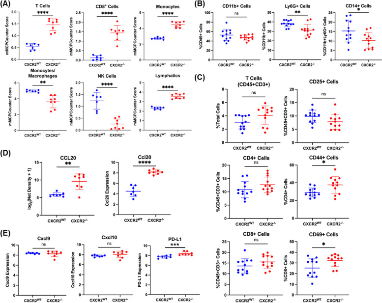
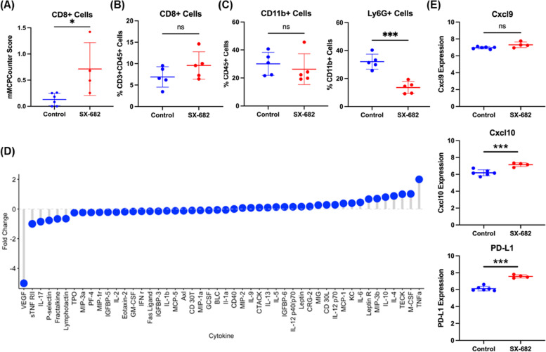
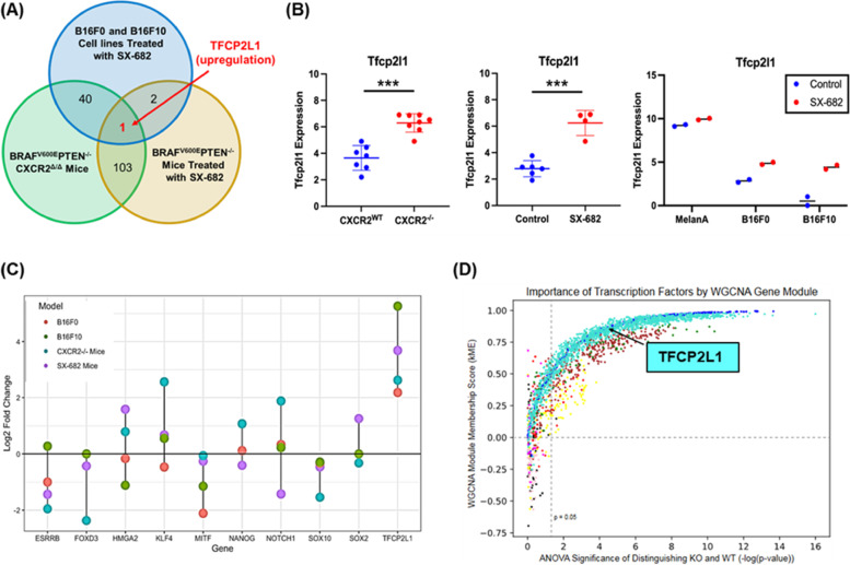
Results: Genetic loss of Cxcr2 or pharmacological inhibition of CXCR1/CXCR2 during melanoma tumor induction resulted in key changes in gene expression that reduced tumor incidence/growth and increased anti-tumor immunity. Interestingly, after Cxcr2 ablation, Tfcp2l1, a key tumor suppressive transcription factor, was the only gene significantly induced with a log2 fold-change greater than 2 in these three different melanoma models.

Conclusions: Here, we provide novel mechanistic insight revealing how loss of Cxcr2 expression/activity in melanoma tumor progenitor cells results in reduced tumor burden and creation of an anti-tumor immune microenvironment. This mechanism entails an increase in expression of the tumor suppressive transcription factor, Tfcp2l1, along with alteration in the expression of genes involved in growth regulation, tumor suppression, stemness, differentiation, and immune modulation. These gene expression changes are coincident with reduction in the activation of key growth regulatory pathways, including AKT and mTOR.

Keywords: CXCR2; Genetic mouse models; Genomic analysis; Melanoma; Tumor immune microenvironment.


Conflict of interest statement

JA Zebala and DY Maeda are affiliated with Syntrix Pharmaceuticals and provided the drug for these studies. The other authors do not have any competing interests to disclose.

Figures

Fig. 1
Fig. 1
CXCR2 is associated with tumorigenesis and poor prognosis. a GEO dataset analysis of expression of CXCR1 and CXCR2 in nevi as compared to melanoma lesions (not significant, Welch's t-test). b GEO dataset analysis of expression of CXCL1, CXCL2, CXCL3, CXCL5 and CXCL8 in nevi and melanoma tissues (significance determined by Welch's t-test). c Overall survival plot of melanoma patients from the TCGA SKCM dataset indicates significantly improved survival (p = 0.035, log-rank test) in the lowest quartile of CXCR2 expression (blue, n = 107) compared to the highest quartile (red, n = 114). d Analysis of survival of 25 melanoma patients treated with anti-PD-1 in relation to high (red) or low (blue) expression of CXCR2 [p < 0.01, log-rank test; [27]]. e Re-analysis of the Riaz RNA-seq database shows CXCR2 expression is lower in melanoma patients who responded to anti-PD1 treatment (p < 0.05, Welch's t-test)
Fig. 2
Fig. 2
CXCR2 knockout decreases melanoma tumor burden. a Tyr-CreER+:: BrafCA/+::Ptenlox4−5/lox4−5::mT/mG C57BL/6 mice were crossed with floxed Cxcr2 mice to obtain mice with inducible tumors with or without CXCR2 expression. Thirty-six days after 4-HT administration, skin tumor volume and count were recorded, and mice were photographed (significance determined by Welch's t-test). Similarly, b Tyr-CreER+::NRasQ61R::Ink4a−/− mice were crossed with floxed Cxcr2 mice, and resulting pups were treated with 4-HT on days 1 and 2 prior to UV irradiation on day 3 to initiate tumor formation (n = 16/genotype). Tumors were measured, counted, and mice were photographed (significance determined by Welch's t-test). RNA was extracted from BrafV600E/Pten−/−/Cxcr2−/− and BrafV600E/Pten−/−/Cxcr2WT tumors and subjected to RNAseq analysis. c A volcano plot showing fold change and significance of differential gene expression in Cxcr2−/− tumors compared to Cxcr2WT tumors. d Gene set enrichment analysis (GSEA) of RNAseq data identifies 8 gene sets enriched in Cxcr2−/− tumors. Point size indicates the gene ratio (percent of genes from the gene set contributing to the enrichment score) and point color represents the FDR q-value
Fig. 3
Fig. 3
The immune infiltrate of BrafV600E/Pten−/− tumors is altered with loss of Cxcr2. a mMCPCounter analysis performed on bulk RNAseq data from BrafV600E/Pten−/−melanoma tumors with or without Cxcr2 predicts significantly enhanced infiltration of T cells, CD8 + T cells, monocytes, NK cells, and lymphatic vessels into Cxcr2−/− tumors. b FACS analysis of CD45 + myeloid cells in BrafV600E/Pten−/− melanoma reveals decreased MDSC-like cells in Cxcr2−/− tumors. c FACS analysis of CD45 + cells in BrafV600E/Pten−/− melanoma tumors identified changes in activated CD4 + CD44 + T cells and CD8 + CD69 + T cells. d Cytokine array for 62 cytokines expressed in TME of BrafV600E/Pten−/− tumors revealed one major cytokine, CCL20, that is strongly upregulated with loss of Cxcr2 (n = 4/genotype) based on net density. These data are complemented by increased Ccl20 mRNA with loss of Cxcr2 in BrafV600E/Pten−/− tumors. e Cxcl9, Cxcl10, and PD-L1 expression based upon RNAseq analysis from BrafV600E/Pten−/− tumors expressing or not expressing Cxcr2 in melanocytes. All statistical significance determined via Welch’s t-test
Fig. 4
Fig. 4
SX-682 affects BrafV600E/Pten−/− and NRasQ61R/Ink4a−/− tumorigenesis. a BrafV600E/Pten−/− and b NRasQ61R/Ink4a−/− mice were fed chow containing SX-682 or vehicle continuously through tumor formation, and tumors were measured and counted. Significance was determined using a Welch's t-test. c A volcano plot showing fold change and significance of differential gene expression between tumors from SX-682-fed and control-fed BrafV600E/Pten−/− mice. d Gene set enrichment analysis of SX-682 treated or control BrafV600E/Pten.−/− tumors identifies gene sets enriched in SX-682 treated tumors (positive normalized enrichment score) or enriched in control tumors (negative normalized enrichment score)
Fig. 5
Fig. 5
SX-682 alters the immune profile of BrafV600E/Pten−/− melanoma. a mMCPCounter analysis of bulk RNAseq data predicts enrichment for CD8 + T cell infiltrate into tumors following treatment with SX-682 (p < 0.05). b FACS analysis confirms a trend toward increased CD8 + T cells in SX-682 treated BrafV600E/Pten−/− melanoma. c FACS analysis of CD45 + myeloid cells indicated a significant decrease in immunosuppressive CD11b + Ly6G + cells, but no change in total CD11b + cells. d A cytokine array was performed on control and SX-682 treated tumors, identifying a notable decrease in Vegf and an increase in Tnfα. e Cxcl9, Cxcl10, and Pd-l1 expression based upon RNAseq analysis from SX-682 or control treated tumors. All statistical significance determined via Welch’s t-test
Fig. 6
Fig. 6
Tumor cell-specific impacts of SX-682. CXCL1 and CXCR2 expression on Melan-A, B16F0, and B16F10 cells based on a the NCBI database and b CXCR2 expression in Melan-A, B16F0 and B16F10 cells based on flow cytometry. c Cell lines were treated with 5 µM SX-682 (or DMSO control) for 4 days prior to staining with Pacific Blue-Ki67 for FACS analysis. The percentage of positive staining cells was significantly decreased in the SX-682 treated cells for all cell lines (analyzed using a two-way ANOVA with Benjamini and Hochberg (BH) correction for multiple tests). d Cytokine array of SX-682-treated Melan-A, B16F0 and B16F10 cells shows that SX-682 treatment reduced the expression of KC and VEGF in all three cell lines
Fig. 7
Fig. 7
Tfcp2l1 is commonly upregulated across three models of CXCR2 perturbation. a, b In comparing expression data from BrafV600E/Pten−/−/Cxcr2−/− tumors. BrafV600E/Pten−/− tumors treated with SX-682, and B16F0 and B16F10 cell lines treated with SX-682, Tfcp2l1 was consistently upregulated compared to appropriate controls (as determined by Welch's t-test). c Log2 fold change for Tfcp2l1 and related genes across experimental groups based upon RNAseq analysis. d Identification of transcription factors central to Weighted Correlation Network Analysis (WGCNA) co-expressed gene modules (by kME) and significantly differentially expressed between BrafV600E/Pten−/−/Cxcr2−/− and BrafV600E/Pten−/−/Cxcr2WT tumors. TFs are colored by gene module and show varying levels of centrality to each module and importance in distinguishing WT and KO tumors. Turquoise dots represent transcription factors that are up in the BrafV600E/Pten−/−/Cxcr2−/− tumors and blue dots represent transcription factors that are up in the BrafV600E/Pten−/−/Cxcr2WT tumors

Update of

References

    1. Hanahan D. Hallmarks of cancer: new dimensions. Cancer Discov. 2022;12(1):31–46. doi: 10.1158/2159-8290.CD-21-1059. - DOI - PubMed
    1. Moser B. Emerging roles of chemokines in cancer immunotherapy. Cancers (Basel) 2022;14(15):3593. doi: 10.3390/cancers14153593. - DOI - PMC - PubMed
    1. Singh S, Nannuru KC, Sadanandam A, Varney ML, Singh RK. CXCR1 and CXCR2 enhances human melanoma tumourigenesis, growth and invasion. Br J Cancer. 2009;100(10):1638–1646. doi: 10.1038/sj.bjc.6605055. - DOI - PMC - PubMed
    1. Vandercappellen J, Van Damme J, Struyf S. The role of CXC chemokines and their receptors in cancer. Cancer Lett. 2008;267(2):226–244. doi: 10.1016/j.canlet.2008.04.050. - DOI - PubMed
    1. Raman D, Sobolik-Delmaire T, Richmond A. Chemokines in health and disease. Exp Cell Res. 2011;317(5):575–589. doi: 10.1016/j.yexcr.2011.01.005. - DOI - PMC - PubMed

Publication types

Substances