. 2023 Jul 11;122(13):2782-2790.
doi: 10.1016/j.bpj.2023.05.034. Epub 2023 Jun 2.

Voxelotor does not inhibit sickle hemoglobin fiber formation upon complete deoxygenation

Affiliations

Voxelotor does not inhibit sickle hemoglobin fiber formation upon complete deoxygenation

Eli H Worth et al. Biophys J. .

Abstract

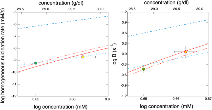
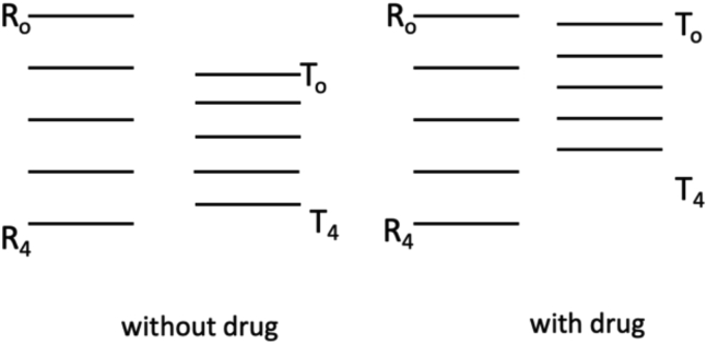
The drug voxelotor (commercially known as Oxbryta) has been approved by the US Food and Drug Administration for the treatment of sickle cell disease. It is known to reduce disease-causing sickling by inhibiting the transformation of the non-polymerizing, high-oxygen-affinity R quaternary structure of sickle hemoglobin into its polymerizing, low-affinity T quaternary structure. It has not been established whether the binding of the drug has anti-sickling effects beyond restricting the change of quaternary structure. By using a laser photolysis method that employs microscope optics, we have determined that fully deoxygenated sickle hemoglobin will assume the T structure. We show that the nucleation rates essential to generate the sickle fibers are not significantly affected by voxelotor. The method employed here should be useful for determining the mechanism of sickling inhibition for proposed drugs.


Conflict of interest statement

Declaration of interests The authors declare no competing interests.

Figures

Figure 1
Figure 1
Hill plot of HbS with and without voxelotor. Y is the fractional saturation with CO, and P is the steady-state laser power, in arbitrary units, which photolyzes the CO. Power was proportional to the output power shown on the laser itself. Each point was measured as an independent experiment in which the laser was turned on for 10 s and absorbance spectra in the Soret region (400–450 nm) were measured before the laser was turned off. The sample was then slightly displaced between points to ensure that each point was fully independent of the others. Voxelotor-modified HbS is shown by the red circles, while unmodified HbS is shown as diamonds. The voxelotor-modified HbS shows a straight line, indicative of a Hb locked in a single quaternary structure over the range of observation. The line also matches the asymptote of the unmodified HbS, which indicates that the R-structure affinity has not been modified with voxelotor. The sample concentration was 29.7 g/dL, measured at 25°C. To see this figure in color, go online.
Figure 2
Figure 2
Progress curves for deoxyHbS polymerization in different multiple small photolysis volumes. 59 spots were illuminated and measured simultaneously, and the procedure was repeated at multiple sites in the sample. (a) Increase in absorbance at 425 nm for a sample of HbS concentration 29.7 g/dL at 25°C in a solution with HbS saturated with voxelotor. The absorbance increases because the concentration gradient of free molecules created by polymerization results in diffusion of molecules into the illuminated spot. Polymerization commences at different times because of the stochastic variation in the homogeneous nucleation of single fibers. When the first fiber forms, subsequent fiber formation occurs by secondary nucleation, explaining the similarity in shape of all of the progress curves. On the axis label, AU stands for absorbance units. (b) Distribution of the tenth times for the experiment shown in (a). The exponential decay rate for the tail of the distribution time is related to the homogeneous nucleation rate (21,27). The solid red curves are a least-squares fit to the data using Szabo’s (27) equation. (c) The distribution of tenth times for a 28.8 g/dL HbS sample without voxelotor. The distributions shown in (b) and (c) decay exponentially past their peaks. That decay rate is directly related to the rate of homogeneous nucleation (27). Following nucleation, each polymerization progress curve grows exponentially at first. To see this figure in color, go online.
Figure 3
Figure 3
Effect of voxelotor on homogeneous and heterogeneous nucleation rates for 29.8-g/dL samples with voxelotor (filled yellow circle) and 28.8 g/dL sample without voxelotor (filled green circle). Horizontal error bars represent our estimated uncertainty in concentraton, primarily based on the spectra fit to the measured data. Vertical error bars are the uncertainty from fitting the specific rate parameters to the data collected. The upper dashed blue lines show the expected values for the rates measured in the presence of 0.055 M sodium dithionite, as done previously. The lower and lighter dashed blue lines are the result of simply displacing the upper dashed lines to match the data. The light dashed red line is the result of using the model for nucleation with a single parameter adjusted to change the contact energy between molecules to match the control sample. The continuous red line shows the same adjustment made to best match both data points. (a) Homogeneous nucleation rates. (b) Exponential growth rate parameter B, which is determined by rates of secondary nucleation and fiber growth (26,43) (cf. materials and methods). In all cases, it is clear that the data with voxelotor present exhibit no decrease in nucleation rates, thus showing that voxelotor’s only effect on fiber formation is to inhibit transformation to the T structure. To see this figure in color, go online.
Figure 4
Figure 4
Free energy allosteric representation of HbS with and without voxelotor. Free energy levels are shown for different ligation states in the two quaternary structures, R and T. In the absence of drug, the switch point is near three ligands. To is more stable than Ro thanks to the presence of several salt bridges. By obstructing two of those, the T state is destabilized, and the entire manifold of T states rises relative to the R states. Consequently, as ligands dissociate in oxygen delivery, voxelotor keeps the molecule in the R state until the final binding step. In the complete absence of ligands, the state To must remain more stable than Ro because only the T structure can polymerize, and our experiments demonstrate the ability of the molecule to polymerize with voxelotor bound.

References

    1. Pauling L., Itano H., et al. Wells I.C. Sickle cell anemia, a molecular disease. Science. 1949;110:543–548. - PubMed
    1. Perutz M.F. Stereochemistry of cooperative effects in haemoglobin. Nature. 1970;228:726–739. http://www.ncbi.nlm.nih.gov/pubmed/5528785 - PubMed
    1. Gelin B.R., Lee A.W., Karplus M. Hemoglobin tertiary structural change on ligand binding. Its role in the co-operative mechanism. J. Mol. Biol. 1983;171:489–559. doi: 10.1016/0022-2836(83)90042-6. https://www.ncbi.nlm.nih.gov/pubmed/6663623 - DOI - PubMed
    1. Padlan E.A., Love W.E. Refined Crystal Structure of Deoxyhemoglobin S I. Restrained lest squares refinement at 3.0 A resolution. J. Biol. Chem. 1985;260:8272–8279. - PubMed
    1. Sunshine H.R., Hofrichter J., et al. Eaton W.A. Oxygen binding by sickle cell hemoglobin polymers. J. Mol. Biol. 1982;158:251–273. - PubMed

MeSH terms

LinkOut - more resources