Review
. 2023 Jun;20(3):194-207.
doi: 10.1007/s11897-023-00600-6. Epub 2023 Jun 5.

Assessment of Right Ventricular Function-a State of the Art

Affiliations
Review

Assessment of Right Ventricular Function-a State of the Art

Abdul Hameed et al. Curr Heart Fail Rep. 2023 Jun.

Abstract

Purpose of review: The right ventricle (RV) has a complex geometry and physiology which is distinct from the left. RV dysfunction and failure can be the aftermath of volume- and/or pressure-loading conditions, as well as myocardial and pericardial diseases.

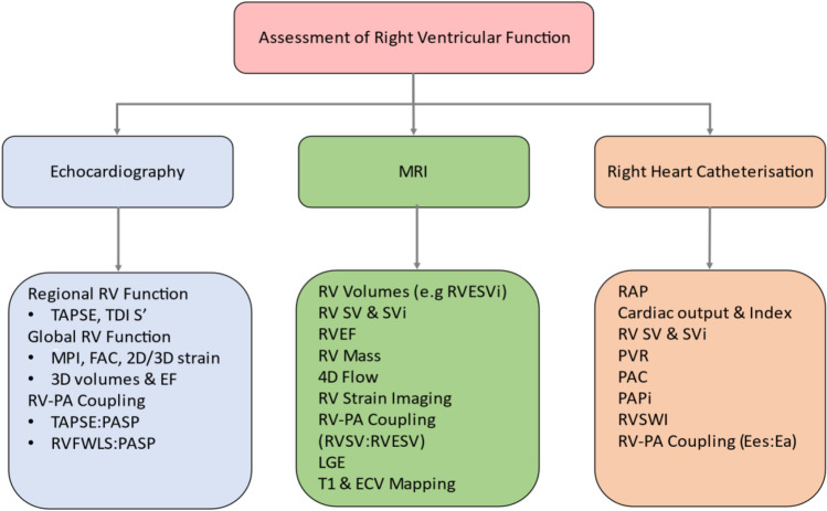
Recent findings: Echocardiography, magnetic resonance imaging and right heart catheterisation can assess RV function by using several qualitative and quantitative parameters. In pulmonary hypertension (PH) in particular, RV function can be impaired and is related to survival. An accurate assessment of RV function is crucial for the early diagnosis and management of these patients. This review focuses on the different modalities and indices used for the evaluation of RV function with an emphasis on PH.

Keywords: Pulmonary hypertension; Right heart catheterisation; Right ventricular ejection fraction; Right ventricular function; Right ventricular-pulmonary arterial coupling.


Conflict of interest statement

A Hameed, R Condliffe, A J Swift, S Alabed, D J Kiely, and A Charalampopoulos declare no conflict of interest.

Figures

Fig. 1
Fig. 1
Echocardiographic parameters for the quantitative assessment of RV function. A Tricuspid annular plane systolic excursion (TAPSE), B right ventricular systolic wave velocity measured with Tissue Doppler Imaging (RV S′), C right ventricular ejection fraction measured by 3D-echocardiography (3DE RVEF), D fractional area change (FAC), E 2D right ventricular global longitudinal strain (GLS). Original image republished without need for permission under a Creative Commons Attribution-Non-Commercial-No Derivatives 4.0 International License from Rana et al. [37]
Fig. 2
Fig. 2
Cardiac MRI cine images (short-axis: left, four chamber: right). Top row: normal MRI. RV; right ventricle, LV; left ventricle, IVS; interventricular septum, RA; right atrium, LA; left atrium. Bottom row: pulmonary arterial hypertension features including a hypertrophied RV myocardium (blue), dilated RV chamber (yellow), IVS straightening (green), RA enlargement (orange) and tricuspid regurgitation jet (arrow)
Fig. 3
Fig. 3
Modalities and parameters used for the assessment of right ventricular function. MRI, magnetic resonance imaging; RV, right ventricular; TAPSE, tricuspid annular plane systolic excursion; TDI S′, right ventricular systolic wave on Tissue Doppler Imaging; MPI, myocardial performance index; FAC, fractional area change; EF, ejection fraction; PA, pulmonary artery; PASP, pulmonary arterial systolic pressure; RVFWLS, right ventricular free wall longitudinal strain; RVESVi, right ventricular end-systolic volume index; SV, stroke volume; SVi, stroke volume index; RVSV, right ventricular stroke volume; RVESV, right ventricular end-systolic volume; LGE, late gadolinium enhancement; ECV, extracellular volume; RAP, right atrial pressure, PVR, pulmonary vascular resistance; PAC, pulmonary arterial compliance; PAPi, pulmonary arterial pulsatility index; RWSVI, right ventricular stroke work index; Ees, end-systolic elastance; Ea, arterial elastance

References

    1. Lippmann MR, Maron BA. The right ventricle: from embryologic development to RV failure. Curr Heart Fail Rep. 2022;19:325–333. doi: 10.1007/s11897-022-00572-z. - DOI - PMC - PubMed
    1. Loukas M, Klaassen Z, Tubbs RS, et al. Anatomical observations of the moderator band. Clin Anat. 2010;23:43–50. doi: 10.1002/ca.20968. - DOI - PubMed
    1. Brown SB, Raina A, Katz D, et al. Longitudinal shortening accounts for the majority of right ventricular contraction and improves after pulmonary vasodilator therapy in normal subjects and patients with pulmonary arterial hypertension. Chest. 2011;140:27–33. doi: 10.1378/chest.10-1136. - DOI - PubMed
    1. Walker LA, Buttrick PM. The right ventricle: biologic insights and response to disease: updated. Curr Cardiol Rev. 2013;9(1):73–81. - PMC - PubMed
    1. Guimaron S, Guihare J, Amsallem M, Haddad F, Fadel E, Mercier O. Current knowledge and recent advances of right ventricular molecular biology and metabolism from congenital heart disease to chronic pulmonary hypertension. Biomed Res Int. 2018;17(2018):1981568). - PMC - PubMed

MeSH terms