

Targeted Knockdown of Macrophage Migration Inhibitory Factor Enhances UVB Irradiation-Induced Apoptosis Via Increasing ROS Generation in Oral Squamous Cell Carcinoma
- PMID: 37272017
- PMCID: PMC10272676
- DOI: 10.1177/15330338231163436
Targeted Knockdown of Macrophage Migration Inhibitory Factor Enhances UVB Irradiation-Induced Apoptosis Via Increasing ROS Generation in Oral Squamous Cell Carcinoma
Abstract
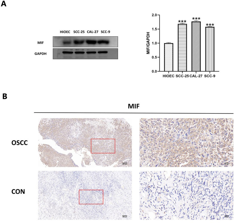
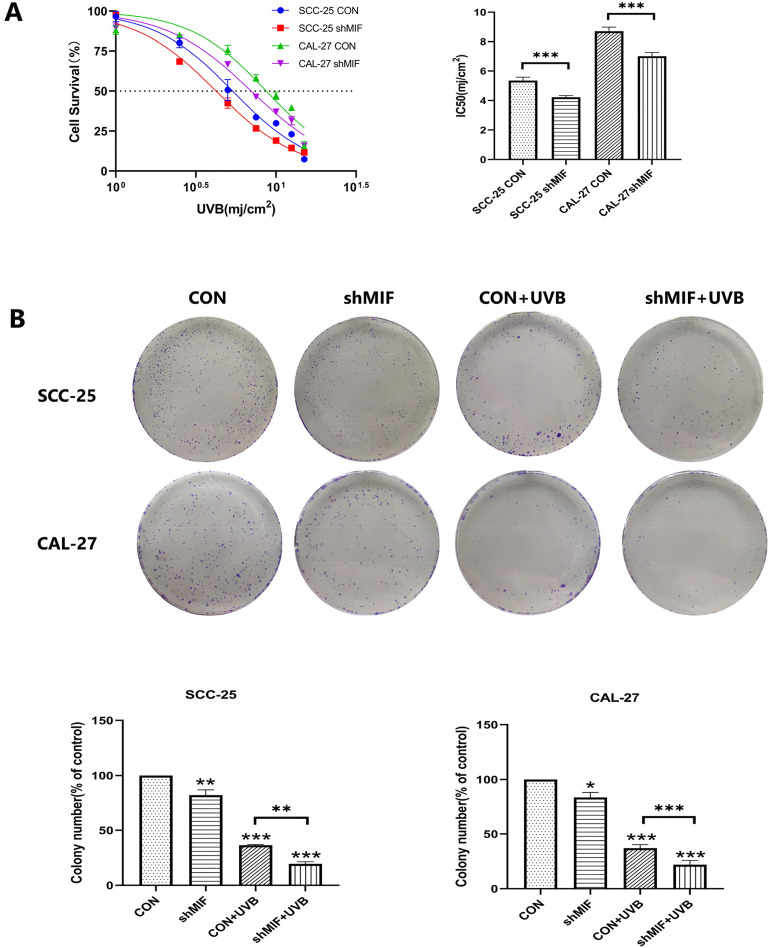
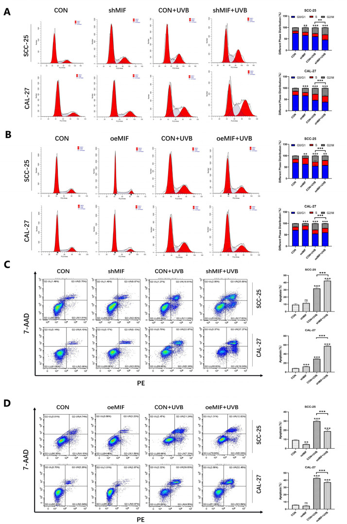
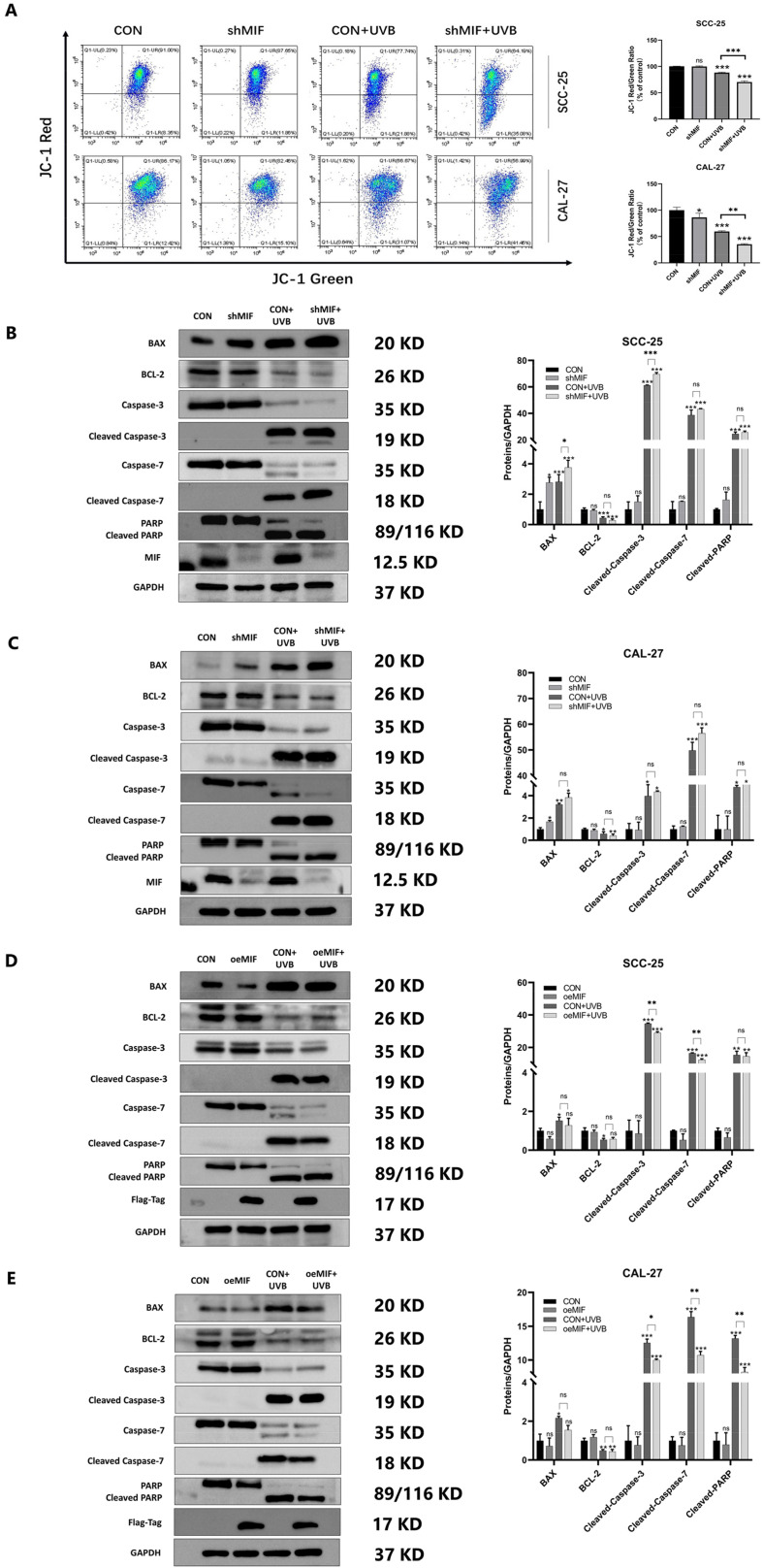
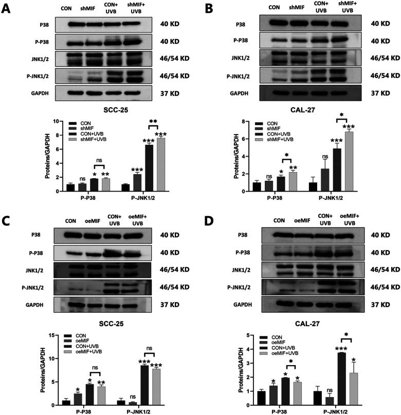
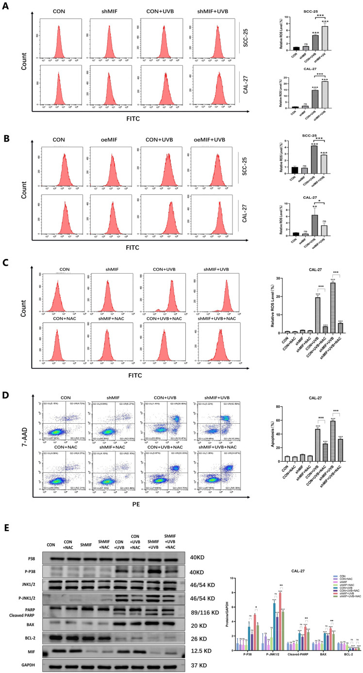
Objectives: We investigated the effects of macrophage migration inhibitory factor (MIF) knockdown or overexpression combined with ultraviolet radiation B (UVB) irradiation on cell proliferation and apoptosis of oral squamous cell carcinoma (OSCC). Methods: MIF expression in OSCC and adjacent tissues was detected by immunohistochemistry. MIF expression in human immortalized oral epithelial cells (HIOEC) and OSCC cells was detected by western blotting. MIF was knocked down or overexpressed in OSCC cell lines (SCC-25 and CAL-27). OSCC cells were set up into control (CON), MIF overexpression/knockdown (oeMIF/shMIF), CON + UVB, and oeMIF + UVB/shMIF + UVB groups based on their exposure to UVB irradiation. Cell line proliferation was studied using a cell counting kit-8 (CCK-8) and colony formation assays. Flow cytometry was applied for determination of apoptosis, cell cycle, reactive oxygen species (ROS) abundance, and mitochondrial membrane potential. Apoptosis-related proteins were assayed by western blotting. Results: The expression of MIF was significantly higher in OSCC tissues and cell lines than in adjacent tissues and HIOEC. MIF knockdown accompanied by UVB irradiation significantly hampered cell viability and proliferation compared to MIF knockdown or UVB irradiation alone. Western blotting and flow cytometry showed that MIF knockdown combined with UVB irradiation not only induced apoptosis via the mitochondrial pathway but also mediated the cell cycle. Flow cytometry showed that ROS and mitochondrial membrane potential depolarization were increased in the combination treatment groups compared with the mono-treatment groups. Additionally, the ROS scavenger N-acetylcysteine significantly attenuated MIF knockdown combined with UVB irradiation-induced apoptosis and reversed MIF knockdown combined with UVB irradiation-induced MAPK activation. Conclusion: MIF knockdown combined with UVB irradiation significantly inhibited the proliferation of OSCC cells. MIF was involved in UVB-induced ROS generation and enhanced UVB irradiation-induced mitochondria-dependent apoptosis of OSCC cells by activating the MAPK pathway. This suggests that MIF-targeted therapy combined with UVB irradiation may be a novel approach for treating OSCC.
Keywords: apoptosis; macrophage migration inhibitory factor; oral squamous cell carcinoma; reactive oxygen species; ultraviolet radiation B irradiation.
Conflict of interest statement
The author(s) declared no potential conflicts of interest with respect to the research, authorship, and/or publication of this article.
Figures
Similar articles
-
Transient transfection of macrophage migration inhibitory factor small interfering RNA disrupts the biological behavior of oral squamous carcinoma cells.Mol Med Rep. 2016 Jan;13(1):174-80. doi: 10.3892/mmr.2015.4525. Epub 2015 Nov 6. Mol Med Rep. 2016. PMID: 26549761 Free PMC article.
-
Eugenol Inhibits the Biological Activities of an Oral Squamous Cell Carcinoma Cell Line SCC9 via Targeting MIF.Anticancer Agents Med Chem. 2022 Aug 4;22(15):2799-2806. doi: 10.2174/1871520622666220324105435. Anticancer Agents Med Chem. 2022. PMID: 35331101
-
AURKA contributes to the progression of oral squamous cell carcinoma (OSCC) through modulating epithelial-to-mesenchymal transition (EMT) and apoptosis via the regulation of ROS.Biochem Biophys Res Commun. 2018 Dec 9;507(1-4):83-90. doi: 10.1016/j.bbrc.2018.10.170. Epub 2018 Nov 16. Biochem Biophys Res Commun. 2018. PMID: 30454901
-
New insights into redox-related risk factors and therapeutic targets in oral squamous cell carcinoma.Oral Oncol. 2023 Dec;147:106573. doi: 10.1016/j.oraloncology.2023.106573. Epub 2023 Nov 9. Oral Oncol. 2023. PMID: 37951115 Review.
-
Biogenic Analysis of the Effect of TERC on Cell Proliferation and Migration of Oral Squamous Cell Carcinoma under Digital Minimally Invasive Treatment.Biomed Res Int. 2022 Aug 18;2022:2102795. doi: 10.1155/2022/2102795. eCollection 2022. Biomed Res Int. 2022. Retraction in: Biomed Res Int. 2023 Jun 21;2023:9798465. doi: 10.1155/2023/9798465. PMID: 36033580 Free PMC article. Retracted. Review.
Cited by
-
Targeting macrophage migration inhibitory factor to inhibit T cell immunosuppression in the tumor microenvironment and improve cancer outcomes in head and neck squamous cell carcinoma.Oral Oncol. 2025 Jan;160:107126. doi: 10.1016/j.oraloncology.2024.107126. Epub 2024 Dec 6. Oral Oncol. 2025. PMID: 39644862
References
Publication types
MeSH terms
Substances
LinkOut - more resources
Full Text Sources
Medical
Research Materials
Miscellaneous
