. 2023 Dec;56(12):e13514.
doi: 10.1111/cpr.13514. Epub 2023 Jun 4.

Insufficient GDF15 expression predisposes women to unexplained recurrent pregnancy loss by impairing extravillous trophoblast invasion

Affiliations

Insufficient GDF15 expression predisposes women to unexplained recurrent pregnancy loss by impairing extravillous trophoblast invasion

Chunzi Lyu et al. Cell Prolif. 2023 Dec.

Abstract

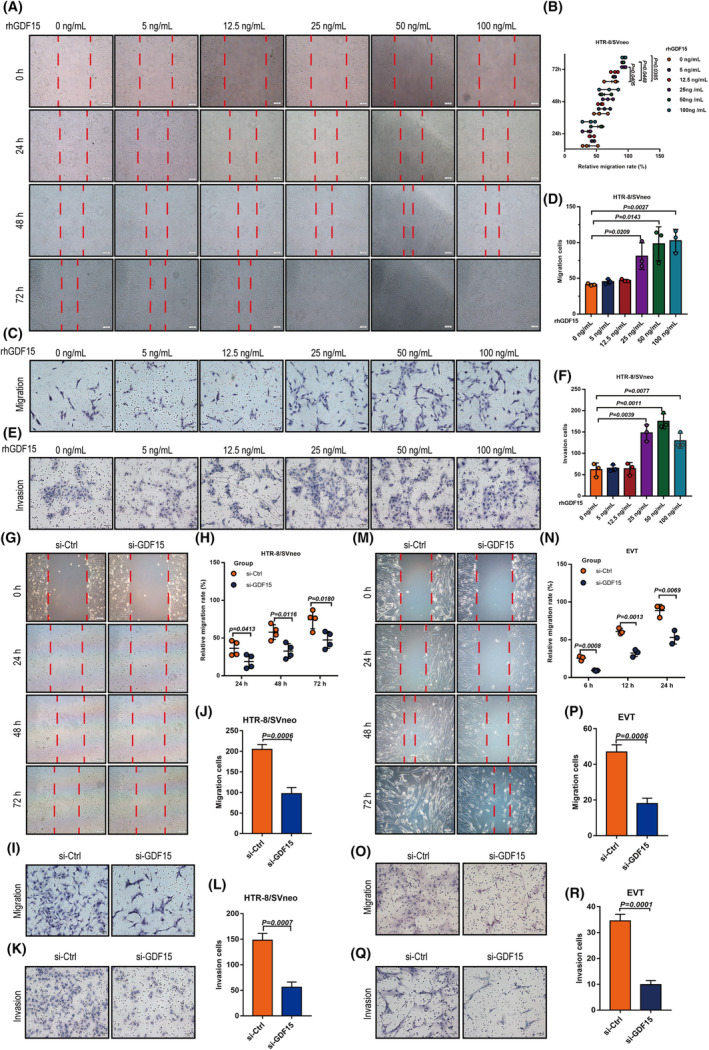
Insufficient extravillous trophoblast (EVT) invasion during early placentation has been shown to contribute to recurrent pregnancy loss (RPL). However, the regulatory factors involved and their involvement in RPL pathogenesis remain unknown. Here, we found aberrantly decreased growth differentiation factor 15 (GDF15) levels in both first-trimester villous and serum samples of unexplained recurrent pregnancy loss (URPL) patients as compared with normal pregnancies. Moreover, GDF15 knockdown significantly reduced the invasiveness of both HTR-8/SVneo cells and primary human EVT cells and suppressed the Jagged-1 (JAG1)/NOTCH3/HES1 pathway activity, and JAG1 overexpression rescued the invasion phenotype of the GDF15 knockdown cells. Induction of a lipopolysaccharide-induced abortion model in mice resulted in significantly reduced GDF15 level in the placenta and serum, as well as increased rates of embryonic resorption, and these effects were reversed by administration of recombinant GDF15. Our study thus demonstrates that insufficient GDF15 level at the first-trimester maternal-foetal interface contribute to the pathogenesis of URPL by impairing EVT invasion and suppressing JAG1/NOTCH3/HES1 pathway activity, and suggests that supplementation with GDF15 could benefit early pregnancy maintenance and reduce the risk of early pregnancy.


Conflict of interest statement

The authors have declared that no conflict of interest exists.

Figures

FIGURE 1
FIGURE 1
GDF15 levels in villi and serum from URPL patients. (A, B) Volcano plot of RNA sequencing data showing the most differentially expressed genes (DEGs) in URPL villi (n = 3) and normal pregnant (control) villi (n = 5). Significantly regulated genes above and below two‐fold are shown in blue and red, respectively. Red dots indicate genes with log2‐fold change >1, blue dots indicate genes with log2‐fold change <1. (C) qPCR was performed to measure the GDF15 mRNA levels in villi from nine URPL patients and nine controls. (D) Western blotting analysis of GDF15 protein levels in villi from six URPL patients and six normal controls. (E) ELISA was performed to analyse serum GDF15 levels in patients with URPL (n = 21) and normal controls (n = 21). (F, G) Haematoxylin and eosin staining and representative immunohistochemical staining against GDF15 in villi and decidua from patients with URPL and normal controls (n = 15/group). EVTs, extravillous trophoblasts; CTBs, cytotrophoblasts; STBs, syncytiotrophoblasts. Data are presented as the means ± SDs. p < 0.01 by two‐tailed Student's t test.
FIGURE 2
FIGURE 2
GDF15 regulates extravillous trophoblast outgrowth in villous explant cultures. (A, B) First‐trimester extravillous explants from normal pregnancies were cultured with 50 ng/mL rhGDF15 for 24 h, and images of the explants were captured under a light microscope after 0 and 24 h. Representative images are shown in the left panel, and summarized quantitative results are shown in the right panel. (C, D) Extravillous explants were transfected with non‐targeting control siRNA (siCtrl) or siRNA targeting GDF15 (si‐GDF15) prior to treatment with or without rhGDF15 (50 ng/mL), and images of the explants were captured under a light microscope after 24 h and 48 h. The migration distance of villus tips was measured and analysed as shown in panel D. (E) Immunofluorescence staining (IF) of GDF15, cytokeratin‐7 and HLA‐G in primary EVTs outgrown from extravillous explants. The levels of GDF15 were assayed by IF (left panel). The knockdown efficiency for GDF15 protein was assessed by ImageJ software (middle panel). The purity of the primary human EVTs was examined by staining cytokeratin‐7 (red) and HLA‐G (green) (right panel). Scale bar, 100 μm. Data are presented as the means ± SD of three independent experiments. p < 0.01 by two‐tailed Student's t test.
FIGURE 3
FIGURE 3
GDF15 promotes invasion and migration of extravillous trophoblasts in vitro. (A, B) HTR‐8/SVneo cells were treated with different concentrations of rhGDF15 (0–100 ng/mL) for cell migration analysis (wound‐healing assays). Representative images are presented in panel A, and the relative migration rate in each group was quantified and summarized in panel B. (C‐D) Transwell™ assays were performed to examine the migratory abilities of rhGDF15‐treated HTR‐8/SVneo cells. Representative images are presented in panel C, and the number of migrating cells in each group was quantified and summarized in panel D. (E, F) Matrigel‐coated Transwell™ assays were performed to study cell invasion in rhGDF15‐treated HTR‐8/SVneo cells. Representative images are presented in panel E, and the number of invading cells in each group was quantified and summarized in panel F. (G, H) HTR‐8/SVneo cell motility was examined with wound‐healing assays after transfection with non‐targeting control siRNA (si‐Ctrl) or siRNA targeting GDF15 (si‐GDF15). Panel G shows representative images of the wound‐healing assay; panel H shows the summarized quantitative results of the wound‐healing assay. (I, J) HTR‐8/SVneo cell motility was examined with Transwell™ assays after transfection with si‐Ctrl or si‐GDF15. (I) The panel shows representative images of the migration assay. (J) The panel shows the summarized quantitative results of the migration assay. (K, L) HTR‐8/SVneo cell invasiveness was examined with Matrigel‐coated Transwell™ assays after transfection with or without si‐GDF15. (K) The panel shows representative images of the invasion assay. (L) The panel shows the summarized quantitative results of the invasion assay. Wound‐healing assay (M, N) and Transwell™ assay (O‐P) were transfected with si‐Ctrl or si‐GDF15. (M) The panel shows representative images of the wound‐healing assay. (N) The panel shows the summarized quantitative results of the wound‐healing assay. (O) The panel shows representative images of the migration assay. (P) The panel shows the summarized quantitative results of the migration assay. (Q‐R) Matrigel‐coated Transwell™ assays were performed to examine cell invasiveness after siRNA‐mediated GDF15 knockdown in primary human EVTs. Representative images are presented in the left panel, and the number of invading cells was quantified and summarized in the right panel. Data are presented as the means ± SDs from at least three independent experiments. p < 0.05, and p < 0.01 by two‐tailed Student's t test.
FIGURE 4
FIGURE 4
JAG1 functions downstream of GDF15 in extravillous trophoblasts. (A) Volcano map of the si‐GDF15‐transfected HTR‐8/SVneo cell transcriptome. GDF15 and JAG1 are labelled. (B) Gene Ontology (GO) term analysis. (C) Kyoto Encyclopedia of Genes and Genomes (KEGG) pathway analysis. (D) qPCR analysis of GDF15, CTNNB1, JAG1, NOTCH1‐3 and HES1 mRNA levels in HTR‐8/SVneo cells transfected with si‐GDF15. (E) Western blotting analysis of GDF15, β‐catenin, JAG1, NOTCH1‐3 and HES1 in HTR‐8/SVneo cells transfected with si‐GDF15. (F) qPCR analysis of GDF15, CTNNB1, JAG1, NOTCH1‐3 and HES1 in primary human EVTs with or without GDF15 knockdown. (G) Western blotting analysis of GDF15, β‐catenin, JAG1, NOTCH3 and HES1 in primary human EVTs transfected with or without si‐GDF15. (H) The association between TCF4 and non‐phosphorylated β‐catenin was assessed by co‐IP analysis with an anti‐non‐phosphorylated β‐catenin antibody in HTR‐8/SVneo cells with or without GDF15 knockdown. (I) qPCR analysis of GDF15, CTNNB1, NOTCH3 and HES1 expression in HTR‐8/SVneo cells with or without JAG1‐expressing adenovirus infection. (J) Western blotting analysis of GDF15, β‐catenin, JAG1, NOTCH3 and HeS1 expression in HTR‐8/SVneo cells with or without JAG1‐expressing adenovirus infection. (K) qPCR analysis showed that JAG1 overexpression increased the mRNA levels of NOTCH3 and HES1 in primary human EVTs. (L) Western blotting analysis showed that JAG1 overexpression increased the protein levels of NOTCH3 and HES1 in primary human EVTs. Data are presented as the means ± SDs from at least three independent experiments. p < 0.05, and p < 0.01 by two‐tailed Student's t test.
FIGURE 5
FIGURE 5
GDF15 promotes invasion and migration of extravillous trophoblasts by upregulating JAG1 expression. (A–F) HTR‐8/SVneo cells were transfected with siRNA targeting GDF15 (si‐GDF15) prior to treatment with or without JAG1 overexpression. Cell mobility was examined with wound‐healing assays (A, B) and Transwell™ assays (C, D); cell invasiveness was examined with Matrigel‐coated Transwell™ assays (E, F). Representative images are presented in panels A, C and E, and the summarized quantitative results are presented in panels B, D and F. (G–L) Primary human EVTs were transfected with siRNA targeting GDF15 (si‐GDF15) prior to treatment with or without JAG1 overexpression, followed by wound‐healing assays (G, H) and Transwell™ assays (I, J) of cell migratory capacity and Matrigel‐coated Transwell™ assays of cell invasiveness (K, L). Representative images are presented in panels G, I and K, and the summarized quantitative results are presented in panels H, J and L. (M) qPCR analysis of GDF15, CTNNB1, JAG1, NOTCH3 and HES1 mRNA levels in HTR‐8/SVneo cells transfected with si‐Ctrl, si‐GDF15 or si‐GDF15 + JAG1‐overexpression vector. (N) Western blotting analysis of GDF15, β‐catenin, JAG1, NOTCH3 and HES1 protein levels in HTR‐8/SVneo cells transfected with si‐Ctrl, si‐GDF15 or si‐GDF15 + JAG1‐overexpression vector. EVTs were transfected with si‐Ctrl, si‐GDF15 or si‐GDF15 + JAG1‐overexpression vector for 48 h. (O) The mRNA levels of GDF15, CTNNB1, JAG1, NOTCH3 and HES1 were assessed by qPCR. (P) The protein expression levels of GDF15, β‐catenin, JAG1, NOTCH3 and HES1 in primary human EVTs were measured by Western blotting. Data are presented as the means ± SDs from at least three independent experiments. p < 0.05, and p < 0.01 by two‐tailed Student's t test.
FIGURE 6
FIGURE 6
Decreased GDF15 level contributes toward the onset of abortion in mice. (A) Wild‐type pregnant mice were treated with LPS (0.25 mg/kg) or rmGDF15 (0.01 mg/kg) at gestational day (GD) 7.5, followed by supplementation with rmGDF15 at GD10.5. (B) GDF15 levels in the serum of mice from different treatment groups (n = 10 mice/group). (C) GDF15 protein levels in murine placentas from different groups (n = 3 mice/group). (D) Gdf15, Jag1, Notch3 and Hes1 mRNA levels in murine placentas from different groups (n = 3 mice/group). (E, F) Embryo resorption rates were examined in different treatment groups of mice with LPS‐induced abortion (n = 10 mice/group). (G, H) Depth of CK7+ trophoblast infiltration into the uterus of pregnant mice from the control, LPS or LPS + rmGDF15 groups (n = 3 mice/group) was observed by haematoxylin–eosin staining or immunofluorescence staining. Relative depth of trophoblast infiltration into the uterus: the ratio of CK7+ trophoblast depth (L1 in red) to the total depth (L2 in orange). Data are presented as the means ± SDs from at least three independent experiments. p < 0.05, and p < 0.01 by two‐tailed Student's t test.
FIGURE 7
FIGURE 7
Schematic illustration indicating how the low embryonic villous GDF15 level increases URPL risk. Insufficient GDF15 expression contributes to the onset of URPL by decreasing EVT invasion via the suppression of JAG1/NOTCH3/HES1 signalling (red arrow). The figure was created with BioRender.com.

References

    1. Practice Committee of the American Society for Reproductive Medicine. Electronic address, a.a.o . Definitions of infertility and recurrent pregnancy loss: a committee opinion. Fertil Steril. 2020;113:533‐535. doi:10.1016/j.fertnstert.2019.11.025 - DOI - PubMed
    1. Dimitriadis E, Menkhorst E, Saito S, Kutteh WH, Brosens JJ. Recurrent pregnancy loss. Nat Rev Dis Primers. 2020;6:98. doi:10.1038/s41572-020-00228-z - DOI - PubMed
    1. ACOG Practice Bulletin No . 200 Summary: Early Pregnancy Loss. Obstet Gynecol. 2018;132:1311‐1313. doi:10.1097/AOG.0000000000002900 - DOI - PubMed
    1. Quenby S, Gallos ID, Dhillon‐Smith RK, et al. Miscarriage matters: the epidemiological, physical, psychological, and economic costs of early pregnancy loss. Lancet. 2021;397:1658‐1667. doi:10.1016/S0140-6736(21)00682-6 - DOI - PubMed
    1. Wang B, Xu T, Li Y, et al. Trophoblast H2S maintains early pregnancy via regulating maternal‐fetal interface immune hemostasis. J Clin Endocrinol Metab. 2020;105:e4275‐e4289. doi:10.1210/clinem/dgaa357 - DOI - PMC - PubMed

Substances