. 2023 May 3;15(5):e38470.
doi: 10.7759/cureus.38470. eCollection 2023 May.

Intrahepatic Glissonean Approach for Robotic Anatomical Liver Resection of Segment 7 Using the Saline-Linked Monopolar Cautery Scissors (SLiC-Scissors) Method: A Technical Case Report With Videos

Affiliations

Intrahepatic Glissonean Approach for Robotic Anatomical Liver Resection of Segment 7 Using the Saline-Linked Monopolar Cautery Scissors (SLiC-Scissors) Method: A Technical Case Report With Videos

Takahisa Fujikawa et al. Cureus. .

Abstract

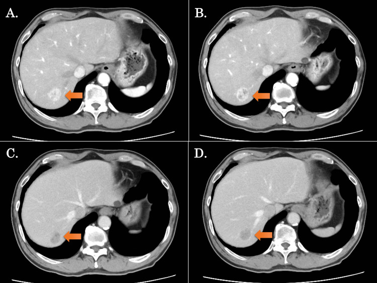
Anatomical hepatectomy of segment 7 (S7) is technically difficult due to its difficult accessibility. Here, we present our experience of robotic anatomical S7 subsectionectomy of the liver employing the saline-linked cautery scissors (SLiC-Scissors) technique. After the right lobe was fully mobilized, dissection of the Glissonean pedicle and hepatic venous branch of S7, as well as the liver parenchymal transection, were safely performed using the SLiC-Scissors method. Despite its technological complexity, the intrahepatic Glissonean approach for robotic anatomical S7 subsectionectomy of the liver employing the SLiC scissors method is safe and efficient.

Keywords: anatomical s7 subsectionectomy; hepatocellular carcinoma; liver parenchymal transection; robotic liver resection; saline-linked cautery scissors.


Conflict of interest statement

The authors have declared that no competing interests exist.

Figures

Figure 1
Figure 1. Preoperative CT images in the present case
A-D show contrast-enhanced CT findings (arrows indicate the tumor); (A, B) The images of the early phase of contrast enhancement, (C, D) Images of the late phase of contrast enhancement. The tumor was located in segment 7, with a maximum diameter of 25 mm.
Figure 2
Figure 2. Trocar placement for robotic anatomical S7 subsectionectomy
Five trocars were typically used for the procedure; robotic #1 port was placed at the right upper lateral side of the abdomen, #3 was placed above the umbilicus, #2 and a 12-mm assistant port were placed between #1 and #3, and #4 was placed at the left-sided epigastrium. The patient cart was rolled in and placed on the right cranial side of the patient. The first assistant surgeon stood on the patient's right side while the second assistant surgeon stood on the left side. T: the targeted lobe of the liver; A: a 12-mm trocar for the assistant surgeon; 1,2,3,4: the four robotic ports; P: Pringle maneuver site
Figure 3
Figure 3. The process of full mobilization of the right liver in robotic anatomical S7 hepatectomy
(A) After the round ligament was severed by laparoscopic coagulating shears, the falciform and right coronary ligaments were dissected using the double bipolar method. (B) The liver was mobilized from the retroperitoneum. (C) A dissection was made between the right adrenal gland and the liver, and the right adrenal vein was clipped and severed. (D) After the right liver was fully mobilized, the Glissonean pedicle in S7, the venous branch in S7, and the hepatic tumor were identified by intraoperative ultrasound and marked.
Figure 4
Figure 4. The process of liver parenchymal transection in robotic anatomical S7 hepatectomy using the SLiC-Scissors method
(A) The root of the G7 was dissected by the water-jet scalpel. (B) G7 was ligated and clipped. (C) The clear demarcation line for S7 was confirmed by intraoperative ICG negative staining. (D) V7 was exposed, clipped, and severed. (E) ICG imaging with the robotic Firefly system was used to correct the displacement of the cut plane as appropriate. (F) After the RHV was exposed from cranially to caudally, liver parenchymal transection was completed. SLiC-Scissors: saline-linked electrocautery scissors, S7: segment 7 of the liver, G7: Glissonean pedicle in S7, ICG: indocyanine green, V7: the venous branch in S7, RHV: right hepatic vein

References

    1. Intrahepatic Glissonean pedicle approach to segment 7 from the dorsal side during laparoscopic anatomic hepatectomy of the cranial part of the right liver. Okuda Y, Honda G, Kobayashi S, Sakamoto K, Homma Y, Honjo M, Doi M. J Am Coll Surg. 2018;226:0–6. - PubMed
    1. Laparoscopic liver resection using the lateral approach from intercostal ports in segments VI, VII, and VIII. Inoue Y, Suzuki Y, Fujii K, et al. J Gastrointest Surg. 2017;21:2135–2143. - PubMed
    1. Laparoscopic liver resection of segment seven: a case report and review of surgical techniques. Takagi K, Kuise T, Umeda Y, Yoshida R, Teraishi F, Yagi T, Fujiwara T. Int J Surg Case Rep. 2020;73:168–171. - PMC - PubMed
    1. A standardized technique for right segmental liver resections. Machado MA, Herman P, Machado MC. Arch Surg. 2003;138:918–920. - PubMed
    1. Laparoscopic liver resection for segment VII lesion using a combination of rubber band retraction method and flexible laparoscope. Lee JW, Choi SH, Kim S, Kwon SW. Surg Endosc. 2020;34:954–960. - PubMed

LinkOut - more resources