

Deep Frontal Lipoma With Frontal Bone Invasion: Report of a Rare Case
- PMID: 37273399
- PMCID: PMC10239272
- DOI: 10.7759/cureus.38546
Deep Frontal Lipoma With Frontal Bone Invasion: Report of a Rare Case
Abstract
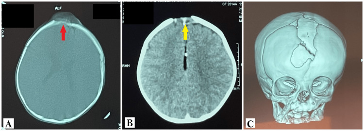
Lipomas are benign masses of fatty tissue, and in the forehead, they may develop in the subcutaneous or deep fat tissue. While subcutaneous lipomas are common, deep forehead lipomas are unusual and rarely invade the underlying bone. Only a few cases have been reported in the literature, and even fewer cases are reported in children. We present a case of a slowly growing frontal mass corresponding to a deep lipoma responsible for frontal bone invasion, resulting in a bony defect reaching the dural space. Through this case, we aim to emphasize forehead lipomas' clinical and surgical characteristics.
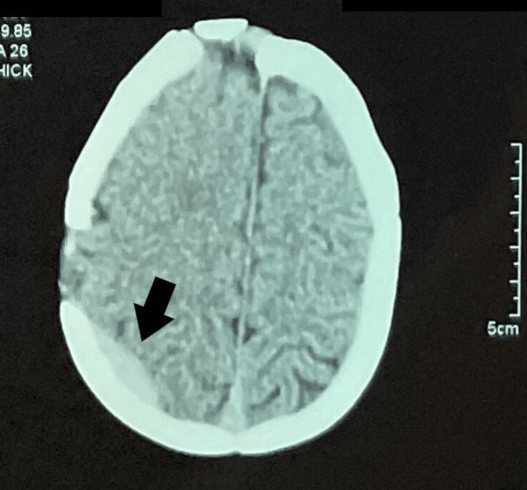
Keywords: bone invasion; extra-dural hematoma; forehead lipoma; frontal lipoma; lipoma.
Copyright © 2023, Ait M'barek et al.
Conflict of interest statement
The authors have declared that no competing interests exist.
Figures

Similar articles
-
Deep forehead lipomas in children-A series and review.Pediatr Dermatol. 2020 May;37(3):520-523. doi: 10.1111/pde.14116. Epub 2020 Feb 25. Pediatr Dermatol. 2020. PMID: 32100337
-
A Case Report of Forehead Subgaleal Lipoma: Diagnostic Dilemmas and Surgical Solutions.Cureus. 2023 Dec 19;15(12):e50760. doi: 10.7759/cureus.50760. eCollection 2023 Dec. Cureus. 2023. PMID: 38239547 Free PMC article.
-
Clinical Characteristics of the Forehead Lipoma.Arch Craniofac Surg. 2014 Dec;15(3):117-120. doi: 10.7181/acfs.2014.15.3.117. Epub 2014 Dec 23. Arch Craniofac Surg. 2014. PMID: 28913203 Free PMC article.
-
Giant Diaphragmatic Lipoma: Two Autopsy Case Reports and Review of the Literature.J Forensic Sci. 2015 Nov;60(6):1640-3. doi: 10.1111/1556-4029.12840. Epub 2015 Aug 10. J Forensic Sci. 2015. PMID: 26258993 Review.
-
Large Intraosseous Lipoma of the Skull: A Case Report and Review of the Literature.World Neurosurg. 2018 Dec;120:525-529. doi: 10.1016/j.wneu.2018.09.149. Epub 2018 Sep 27. World Neurosurg. 2018. PMID: 30268544 Review.
References
-
- Subgaleal lipomas. Zitelli JA. Arch Dermatol. 1989;125:384–385. - PubMed
-
- Frontalis-associated lipoma of the forehead. Salasche SJ, McCollough ML, Angeloni VL, et al. J Am Acad Dermatol. 1989;20:462–468. - PubMed
-
- Subaponeurotic lipoma of the forehead. Grosshans E, Fersing J, Marescaux J. https://pubmed.ncbi.nlm.nih.gov/3605964/ Ann Dermatol Venereol. 1987;114:335–340. - PubMed
-
- Lipoma of the oral and maxillofacial region: site and subclassification of 125 cases. Furlong MA, Fanburg-Smith JC, Childers EL. Oral Surg Oral Med Oral Pathol Oral Radiol Endod. 2004;98:441–450. - PubMed
-
- Lipomas of the oral cavity: clinical findings, histological classification and proliferative activity of 46 cases. Fregnani ER, Pires FR, Falzoni R, Lopes MA, Vargas PA. Int J Oral Maxillofac Surg. 2003;32:49–53. - PubMed
Publication types
LinkOut - more resources
Full Text Sources