

Outcome of the Masquelet Technique for Complex Bilateral Distal Femoral Bone Defects
- PMID: 37273406
- PMCID: PMC10238076
- DOI: 10.7759/cureus.38503
Outcome of the Masquelet Technique for Complex Bilateral Distal Femoral Bone Defects
Abstract
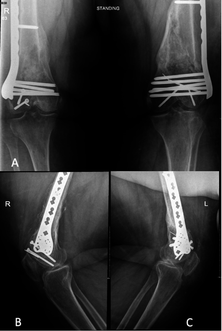
Bone defects are severe conditions caused by various etiologies, including trauma, tumor resection, or chronic osteomyelitis. Different surgical interventions can be utilized to manage such cases, including autologous graft or allograft implantation, distraction osteogenesis, acute shortening, amputation, or the induced membrane technique. Herein, the case of a 39-year-old woman with complex bilateral distal femoral fractures with intra-articular extension is presented. The fractures were accompanied by a significant metaphyseal bone defect, which was managed successfully using the induced membrane Masquelet technique. The patient fully healed despite residual knee joint contractures that did not inhibit her mobility. In conclusion, the Masquelet technique successfully manages complex bone defects and restores functionality even in bilateral simultaneous open bone defects.
Keywords: bilateral distal femur; bone defect; case report; intraarticular fractures; masquelet technique; mva (motor vehicle accident).
Copyright © 2023, Aljaafri et al.
Conflict of interest statement
The authors have declared that no competing interests exist.
Figures


Similar articles
-
[Masquelet technique for the treatment of large dia- and metaphyseal bone defects].Oper Orthop Traumatol. 2015 Aug;27(4):357-68. doi: 10.1007/s00064-014-0300-9. Epub 2015 May 29. Oper Orthop Traumatol. 2015. PMID: 26018724 German.
-
Restoration of long bone defects treated with the induced membrane technique: protocol and outcomes.Injury. 2016 Dec;47 Suppl 6:S53-S61. doi: 10.1016/S0020-1383(16)30840-3. Injury. 2016. PMID: 28040088
-
Reconstruction of a tibial diaphyseal bone defect using the Masquelet technique. A case report.Trauma Case Rep. 2022 Nov 2;42:100728. doi: 10.1016/j.tcr.2022.100728. eCollection 2022 Dec. Trauma Case Rep. 2022. PMID: 36386425 Free PMC article.
-
[Biological reconstruction of large bone defects : Masquelet technique and new procedures].Unfallchirurgie (Heidelb). 2023 Mar;126(3):184-189. doi: 10.1007/s00113-022-01267-9. Epub 2022 Dec 27. Unfallchirurgie (Heidelb). 2023. PMID: 36573997 Review. German.
-
Induced Membrane Technique (Masquelet) for Bone Defects in the Distal Tibia, Foot, and Ankle: Systematic Review, Case Presentations, Tips, and Techniques.Foot Ankle Clin. 2020 Dec;25(4):537-586. doi: 10.1016/j.fcl.2020.08.013. Foot Ankle Clin. 2020. PMID: 33543716
References
-
- Current management of long bone large segmental defects. Lasanianos NG, Kanakaris NK, Giannoudis PV. Orthop Trauma. 2010;24:149–163.
-
- Management of segmental bone defects. [ May; 2022 ];Mauffrey C, Barlow BT, Smith W. J Am Acad Orthop Surg. 2015 23:143–153. - PubMed
-
- The concept of induced membrane for reconstruction of long bone defects. Masquelet AC, Begue T. Orthop Clin North Am. 2010;41:27–37. - PubMed
-
- Reconstruction of the long bones by the induced membrane and spongy autograft [Article in French] Masquelet AC, Fitoussi F, Begue T, Muller GP. http://10929461. Ann Chir Plast Esthet. 2000;45:346–353. - PubMed
Publication types
LinkOut - more resources
Full Text Sources