Case Reports
. 2023 May 31;18(8):2697-2703.
doi: 10.1016/j.radcr.2023.05.028. eCollection 2023 Aug.

A rare case of a primary central nervous system neuroblastoma mimicking a trigeminal schwannoma in an adult

Affiliations
Case Reports

A rare case of a primary central nervous system neuroblastoma mimicking a trigeminal schwannoma in an adult

Bui Quang Huynh et al. Radiol Case Rep. .

Abstract

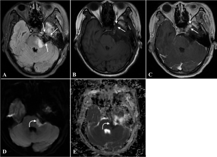
Neuroblastoma is a malignant extra-cranial tumor that frequently arises in the pediatric population aged <5 years but is rare in adults. Only a few cases of primary central nervous system neuroblastoma (PCN-NB) have been documented, with most occurring in young patients. In this article, we report an adult case with a PCN-NB in the cerebellopontine angle-middle cranial fossa region that mimicked another neoplasm. We also discuss the magnetic resonance imaging features and pathological characteristics of PCN-NB and differential diagnosis strategies.

Keywords: Cerebellopontine angle; Primary central nervous system neuroblastoma; Trigeminal schwannoma.


Figures

Fig 1
Fig. 1
The preoperative brain MRI. It showed a left cerebellopontine angle extra-cranial lesion extending into Meckel's cave and the middle cranial fossa. (B) Peripheral hyperintensity and central hypointensity in axial postcontrast T1W axial images corresponding to peripheral hypointensity and central hyperintensity in T2W (A) axial and (D) coronal images (straight arrow). (C) Postgadolinium T1W images show heterogeneous contrast enhancement with a solid enhancing component in the middle cranial fossa. Heterogeneous diffusion restriction areas are observed on the (E) DWI and (F) ADC map (curved arrow).
Fig 2
Fig. 2
MRI brain images at 4 days postsurgery. (A) An axial fluid-attenuated inversion recovery image shows an enlargement of the subarachnoid space in the left cerebellopontine angle (straight arrows). Comparison of (B) pre- and (C) postcontrast T1W images shows enhancing residual tumor in lateral cavernous sinus wall (straight arrows). (D) An axial DWI with corresponding (E) ADC map shows diffusion restriction in the left middle cerebellar peduncle and pons due to cerebral ischemia adjacent to the tumor bed area (curved arrows).
Fig 3
Fig. 3
Histopathological images. (A) A microscopic histopathological image showing small round tumor cells arranged in sheets dispersed in the stroma (PAS staining; ×200 magnification). Immunohistochemical staining of tumor cells (x200 magnification) for (B) OLIG2, (C) CD99, (D) NESTIN, (E) SYNAP, (F) CK, and (G) CHRO.
Fig 4
Fig. 4
MRI brain images at 55 days postsurgery. (A) An axial precontrast T1W image showing a rapidly growing recurrent tumor in the left cerebellopontine angle (straight arrows) spreading through Meckel's cave and extending into the left middle cranial fossa, invading the cavernous sinus and surrounding the left internal carotid artery (curved arrow). The cerebellar peduncle, brainstem, and fourth ventricle were compressed and displaced to the right. (B) A postcontrast T1W image showing a contrast-enhancing lesion. The lesion with diffusion restriction appears (C) bright on DWIs and (D) dark on the ADC map.

References

    1. Borni M, Znazen M, Mdhaffar N, Boudawara MZ. A rare case of pediatric primary central nervous system differentiating neuroblastoma: an unusual and rare intracranial primitive neuroectodermal tumor (a case report) Pan Afr Med J. 2021;40:33. doi: 10.11604/pamj.2021.40.33.30587. - DOI - PMC - PubMed
    1. Lu X, Zhang X, Deng X, Yang Z, Shen X, Sheng H, et al. Incidence, treatment, and survival in primary central nervous system neuroblastoma. World Neurosurg. 2020;140:e61–e72. doi: 10.1016/j.wneu.2020.04.145. - DOI - PubMed
    1. Louis DN, Perry A, Reifenberger G, von Deimling A, Figarella-Branger D, Cavenee WK, et al. The 2016 World Health Organization Classification of Tumors of the Central Nervous System: a summary. Acta Neuropathol. 2016;131(6):803–820. doi: 10.1007/s00401-016-1545-1. - DOI - PubMed
    1. Hart MN, Earle KM. Primitive neuroectodermal tumors of the brain in children. Cancer. 1973;32(4):890–897. doi: 10.1002/1097-0142(197310)32:4<890::aid-cncr2820320421>3.0.co;2-o. - DOI - PubMed
    1. Horten BC, Rubinstein LJ. Primary cerebral neuroblastoma. A clinicopathological study of 35 cases. Brain. 1976;99(4):735–756. doi: 10.1093/brain/99.4.735. - DOI - PubMed

Publication types

LinkOut - more resources