Review
. 2023 May 18:20:100672.
doi: 10.1016/j.mtbio.2023.100672. eCollection 2023 Jun.

Multifunctional nanostructures: Intelligent design to overcome biological barriers

Affiliations
Review

Multifunctional nanostructures: Intelligent design to overcome biological barriers

Mehdi Azizi et al. Mater Today Bio. .

Abstract

Over the past three decades, nanoscience has offered a unique solution for reducing the systemic toxicity of chemotherapy drugs and for increasing drug therapeutic efficiency. However, the poor accumulation and pharmacokinetics of nanoparticles are some of the key reasons for their slow translation into the clinic. The is intimately linked to the non-biological nature of nanoparticles and the aberrant features of solid cancer, which together significantly compromise nanoparticle delivery. New findings on the unique properties of tumors and their interactions with nanoparticles and the human body suggest that, contrary to what was long-believed, tumor features may be more mirage than miracle, as the enhanced permeability and retention based efficacy is estimated to be as low as 1%. In this review, we highlight the current barriers and available solutions to pave the way for approved nanoformulations. Furthermore, we aim to discuss the main solutions to solve inefficient drug delivery with the use of nanobioengineering of nanocarriers and the tumor environment. Finally, we will discuss the suggested strategies to overcome two or more biological barriers with one nanocarrier. The variety of design formats, applications and implications of each of these methods will also be evaluated.

Keywords: Biological barriers; Biotechnology; Cancer; Nanoparticles engineering; Nanotechnology; Targeted drug delivery.


Conflict of interest statement

The authors declare that they have no known competing financial interests or personal relationships that could have appeared to influence the work reported in this paper.

Figures

Image 1
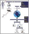
Graphical abstract
Fig. 1
Fig. 1
Four major levels of biological barriers. (1) intravascular barriers, (2) endothelial barriers, (3) extracellular barriers, and (4) cellular barriers. Depending on the depth of the target region, the number of biological barriers differs. Organs are easy sites to deliver nanodrugs, while subcellular organelles are difficult because nanodrugs must cross more barriers to reach the targeted region.
Fig. 2
Fig. 2
Artificial RBCs enable longer circulation of NPs. (A) Construction and properties of resultant RRBC, (B) (a–c), Circulation, oxygen delivery potential and other biomedical application. Reprinted with permission [23]. Copyright 2020, American Chemical Society.
Fig. 3
Fig. 3
FUS mechanism of action for improving EPR in solid tumors. (A) Real macroscopic images of glioma tumors showing FUS-mediated disruption of the BBB, which enhances drug extravasation in BT474-Gluc metastatic brain tumors. Reprinted with permission [83]. Copyright 2018, National Acad Sciences (B) Cartoon animation of the combined work of US and NO therapy results in effective vascular barrier disruption for NP delivery. Reprinted with permission [84]. Copyright 2017, Elsevier.
Fig. 4
Fig. 4
Tumor vasculature normalization to cross the vascular barrier. (A) Functional and structural changes in TEC markers occur during vascular normalization to promote enhanced blood perfusion in tumors for drug delivery. Reprinted with permission [92]. Copyright 2016, Elsevier. (B) Vascular normalization properties of Au NPs can inhibit tumor metastasis by closing vascular gaps. Reprinted with permission [93]. Copyright 2020, American Chemical Society.
Fig. 5
Fig. 5
Tumor vascular disruption for enhanced NP delivery to tumor cells. The preparation and mechanism of action of Gel/CA4−MP/DTX for sequential drug delivery and locally synergistic chemotherapy of osteosarcoma are depicted. Reprinted with permission [38]. Copyright 2018, Elsevier.
Fig. 6
Fig. 6
Tumor vascular infarction. (A) Schematic of encapsulating thrombin and Dox and making chitosan-based polymer NPs. After ionic gelation, NPs were surface functionalized with sulfo-SMCC to supply the maleimide group for the Michael addition process with the CREKA peptide. (B)NP activity in the tumor. When CREKA contacts fibrin-fibronectin complexes inside tumor arteries, the NPs release thrombin to create a thrombus, while Dox (black dots) kills tumor cells. Reprinted with permission [101]. Copyright 2020, Nature Publication Groups.
Fig. 7
Fig. 7
Tumor deoxygenation for starving tumor cells. MS NPs are intratumorally injected for cancer-starving therapy. The tumor blood capillaries produce in situ SiO2 blockers to deoxygenate tissue and prevent reoxygenation after the acidic tumor microenvironment activates NPs. Deoxygenated tumors suffocate without energy metabolism. (MS NPs are PVP-modified). Reprinted with permission [40]. Copyright 2017, Nature Publication Groups.
Fig. 8
Fig. 8
Enzyme-based controlled charge-changing principle for prompting active transport of nanomedicines through trans-endothelial and transcellular barriers. (A) Schematics of working principle. (B) Cationization reaction of enzyme-activatable polymer–drug. Reprinted with permission [103]. Copyright 2019, Nature Publication Groups.
Fig. 9
Fig. 9
Trojan system for crossing the vascular barrier and NP delivery. Making LD-MDS, turning it into nanovesicles with legumain protease, and targeting it for lung metastases. LD-MDS preferentially are applied for lung metastases and converts into nanovesicles and secondary nanovesicles for successful antimetastasis therapy. Reprinted with permission [112]. Copyright 2018, American Chemical Society.
Fig. 10
Fig. 10
Neutrophils are Trojan systems for the tumor-targeted delivery and localization of NPs. In vivo, cabolantinib increases neutrophil-associated, dye-loaded BSA-NPs in prostate cancers. Fluorescent images were overlaid with bright field images used for treatment groups (A) BSA-NP, (B) cabozantinib ​+ ​uncoated NP, (C) BSA-NP, and (D) BSA-NP ​+ ​Ly6G antibody (neutrophil depletion). (E) Tumors were identified on superimposed fluorescence/bright field images using ImageJ, and the mean fluorescence was calculated to estimate NP uptake. Reprinted with permission [117]. Copyright 2021, American Association for Cancer Research.
Fig. 11
Fig. 11
Stroma cell reeducation strategy for ECM remodeling of desmoplastic tumors. (a) Schematic of pH-responsive gold NPs-based system for co-delivery ATRA and HSP47 siRNA. Anionic ATRA and PEG-grafted polyethyleneimine (PEI)-coated gold nanoparticles electrostatically adsorbed siRNA. (b) Schematic of nanosystem stroma modulation and PSC re-education. In the acidic pancreatic tumor microenvironment (pHe 6.5), the nanosystem “activates” (PEG shedding, size decrease, charge rise, and hydrophobic ligand exposure) and exhibits pHe and ATRA dual-enhanced cellular uptake and HSP47 knockdown in PSCs. The desmoplastic stroma is homeostatically repaired, blood perfusion and medication delivery improve, and activated PSCs become quiet. Reprinted with permission [132]. Copyright 2018, Nature Publication Groups. (For interpretation of the references to colour in this figure legend, the reader is referred to the Web version of this article.)
Fig. 12
Fig. 12
Sperm-bioinspired microswimmer for tumor cell internalization (A) design concept. (B) SEM images of 3D-printed tetrapod microstructures. (C) (a) Illustrations of the working principle for the release of drug-loaded sperm on a microfluidic chip. (b) Sperm liberation process upon hitting arms with HeLa spheroids. (c) Distribution of DOX-HCl in HeLa cells. (d) SEM images showing HeLa cell fusion with (i) DOX-HCl-loaded sperm or (ii) unloaded sperm. Live cells are shown with blue arrows, while red arrows indicate apoptotic cells. Reprinted with permission [141]. Copyright 2018, American Chemical Society. (For interpretation of the references to colour in this figure legend, the reader is referred to the Web version of this article.)
Fig. 13
Fig. 13
Schematics of nanobombs for crossing the ECM barrier. (A) Example of light-triggered decomposition of pH-assembled nanobombs in tumors. Reprinted with permission [143]. Copyright 2017, John Wiley and Sons, Inc. (B) Dendrimer-based cluster nanobombs with instantaneous size switching in a tumor acidic environment enable improved tumor penetration. Reprinted with permission [148]. Copyright 2016, National Academy of Science.
Fig. 14
Fig. 14
Orthotopic Pancreatic Cancer Treatment with Polymer-Peptide Conjugates: Ultrasound-Activated Cascade Effect (A) PTPK molecular structure and US activation cascade. (B) Synergistic PTPK internalization and US-induced cascade impact limit tumor development. Reprinted with permission [152]. Copyright 2020, Elsevier.
Fig. 15
Fig. 15
NP surface charge for selective targeting of cancer cell organelle. (A) Positively charged (blue) and negatively charged (red) ligands make up the NP design. (B) The idea behind and the method by which mixed-charge NPs crystallize in cancer lysosomes and destroy cancer cells in a targeted manner. Effectiveness against various cancer cells. Reprinted with permission [163]. Copyright 2020, Nature Publication Groups. (For interpretation of the references to colour in this figure legend, the reader is referred to the Web version of this article.)
Fig. 16
Fig. 16
Logic gate activation of a DNA nanorobot (oncotropic vector) allows selective recognition of different types of tumors. I). Front orthographic schematic of a protein-carrying closed nanorobot (A). Left (boxed) and right DNA-aptamer locks safeguard the device's front. The aptamer lock mechanism is a blue DNA aptamer and a partially complementary strand (orange). Lock antigen keys can keep it separated (red). The lock duplex is always 24 bp, while the non-aptamer strand has an 18–24 base thymine spacer. (C) A protein-released nanorobot. Back scaffold hinges separate blue and orange domains. II) Logic-gated nanorobot activation of different cell types. Nanorobots carry fluorescently tagged HLA A/B/C-specific antibody Fab’ fragments. Nanorobots bind to unlocked HLA-A/B/C-expressing cells (top). Key cells (middle) keep them quiescent, but key ​+ ​cells activate them (bottom). Nanorobot activation truth table. Cell-expressed chemical keys activate the AND gates in aptamer-encoded locks. Colors like a lock and key. Nanorobot conformation is output from aptamer-antigen activation. (C) Eight nanorobots (10–100 ​fmol, loaded with anti-HLA-A/B/C antibody fragments at a molar excess of 20) were tested with six cell types expressing various antigen key combinations for 5 ​h. Each histogram displays cell number versus fluorescence from anti-HLA-A/B/C tagging. Reprinted with permission [165]. Copyright 2012, American Association for the Advancement of Science. (For interpretation of the references to colour in this figure legend, the reader is referred to the Web version of this article.)
Fig. 17
Fig. 17
Schematic of All of the biological barriers to targeted delivery of NPs. The body eliminates most of the NPs before it reaches the targeted region.
Fig. 18
Fig. 18
Customized medication NPs. (A) Samples of the solid tumor are biobank. Slides of solid tumors (avatar animal/patient-derived xerographs, or PDX) are explanted into immunocompromised mice. After three passes, patient-derived cell line cultures (PDCCs) are injected into mice to make drug-testing patient-derived cell xerographs (PDCXs). (B) Single-cell techniques and spatiotemporal heterogeneity imaging are utilized in parallel to assess tumor specimen Eco/Evo heterogeneity. (C) Additional field professionals analyze data. (D) Additional NPs tailoring based on tumor characterization and NP pharmacodynamic and pharmacokinetic predictions based on tumor tissue features. (E) Nanoparticles are tested in vitro and silico, and if early results are positive (F), clinical testing is done.

References

    1. Baghban R., Roshangar L., Jahanban-Esfahlan R., Seidi K., Ebrahimi-Kalan A., Jaymand M., Kolahian S., Javaheri T., Zare P. Cell Commun. Signal. 2020;18:1–19. - PMC - PubMed
    1. Jahanban-Esfahlan R., de la Guardia M., Ahmadi D., Yousefi B. J. Cell. Physiol. 2018;233:2019–2031. - PubMed
    1. Jahanban-Esfahlan R., Seidi K., Zarghami N. Int. J. Hematol. 2017;105:244–256. - PubMed
    1. Stylianopoulos T., Munn L.L., Jain R.K. Trends in cancer. 2018;4:258–259. - PMC - PubMed
    1. Cheng Y.-H., He C., Riviere J.E., Monteiro-Riviere N.A., Lin Z. ACS Nano. 2020;14:3075–3095. - PMC - PubMed

LinkOut - more resources