Review
. 2022 Dec 23;7(3):100129.
doi: 10.1016/j.shj.2022.100129. eCollection 2023 May.

Secondary Mitral Regurgitation: Cardiac Remodeling, Diagnosis, and Management

Affiliations
Review

Secondary Mitral Regurgitation: Cardiac Remodeling, Diagnosis, and Management

Alex L Huang et al. Struct Heart. .

Abstract

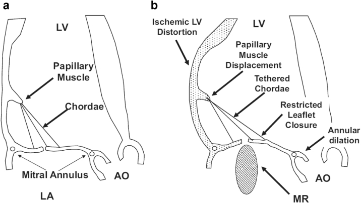
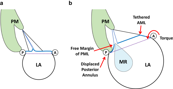
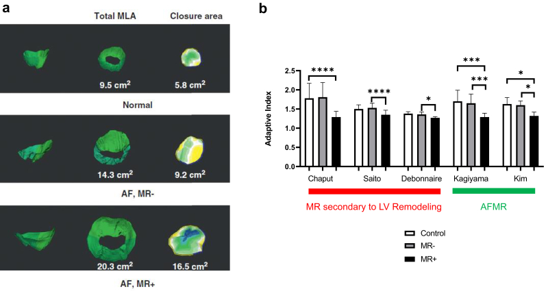
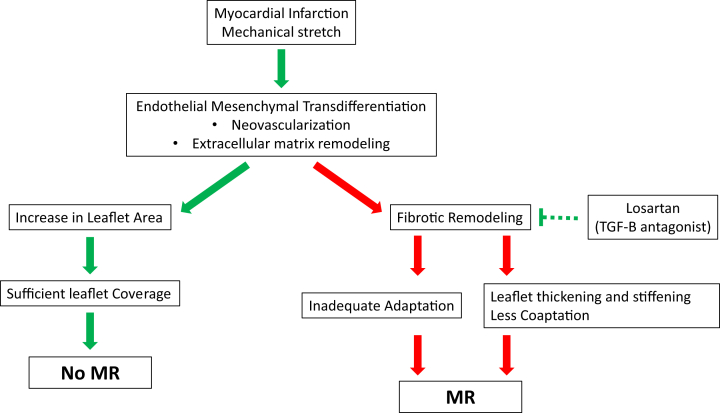
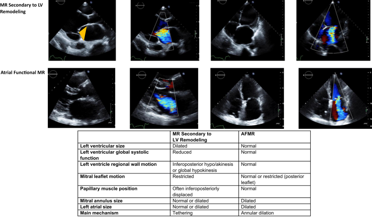
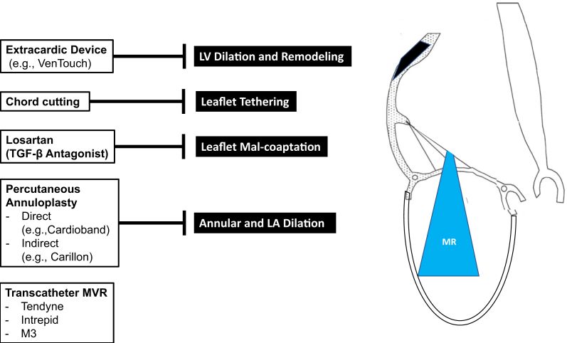
Secondary mitral regurgitation (MR) refers to MR resulting from left ventricular or left atrial remodeling. In ischemic or nonischemic cardiomyopathy, left ventricular dilation (regional or global) leads to papillary muscle displacement, tethering, and leaflet malcoaptation. In atrial functional MR, MR occurs in patients with left atrial dilation and altered mitral annular geometry due to atrial fibrillation. In addition to cardiac remodeling, leaflet remodeling is increasingly recognized. Mitral leaflet tissue actively adapts through leaflet growth to ensure adequate coaptation. Leaflets, however, can also undergo maladaptive thickening and fibrosis, leading to increased stiffness. The balance of cardiac and leaflet remodeling is a key determinant in the development of secondary MR. Clinical management starts with detection, severity grading, and identification of the underlying mechanism, which relies heavily on echocardiography. Treatment of secondary MR consists of guideline-directed medical therapy, surgical repair or replacement, and transcatheter edge-to-edge repair. Based on a better understanding of pathophysiology, novel percutaneous mitral repair and replacement devices have been developed and clinical trials are underway.

Keywords: Echocardiography; Heart failure; Mitral regurgitation.


Figures

Figure 1
Figure 1
Mitral valve anatomy in (a) long axis view and (b) short axis view. (c) Schematic diagram showing the saddle-shape mitral annulus. The mitral apparatus is a complex structure consisting of the annulus, leaflets, chordae tendineae, and the papillary muscles. The annulus is bound by the fibrous trigones and in continuity with the noncoronary cusps and left coronary cusps of the aortic valve. The mitral annulus has a saddle shape with high points anteriorly and posteriorly, and low (=ventricular) points lateral-medially. Modified from Otto CM and Carpentier A.
Figure 2
Figure 2
Mechanism of ischemic mitral regurgitation. (a) Normal left ventricular and mitral valve geometry. (b) In ischemic MR, LV remodeling leads to LV apical displacement of papillary muscles, mitral leaflet tethering, and restricted leaflet closure. Adopted from Hung JW. Abbreviations: AO, aorta; LA, left atrium; LV, left ventricle; MR, mitral regurgitation.
Figure 3
Figure 3
Proposed mechanism of atrial functional mitral regurgitation (AML). (a) Normal left atrial and mitral annulus geometry. (b) In atrial mitral regurgitation, left atrial dilation pulls the mitral annulus posteriorly onto the crest of the LV inlet. This reduces the free edge/area of the posterior leaflet. The annulus displaces superiorly and “flattens” the anterior leaflet. This results in an increased distance between the annulus and papillary muscles, leading to restricted leaflet motion during systole. Modified from Silbiger JJ. Abbreviations: AML, anterior mitral leaflet; LA, left atrium; LV, left ventricle; MR, mitral regurgitation; PM, papillary muscle; PML, posterior mitral leaflet.
Figure 4
Figure 4
Leaflet adaptation. (a) Three-dimensional echocardiography can measure total MV leaflet and mitral annular closure area. Compared to control, patients with AF, even without mitral regurgitation (AF, MR−) have a bigger average leaflet area. However, those that develop atrial functional mitral regurgitation (AF, MR+) also have a bigger annular closure area. Thus, despite an increase in total leaflet area, there is a leaflet to annular area mismatch, resulting in mitral regurgitation. Figure adopted from Kim et al. (b) Studies,,, , on leaflet adaptation in MR. Adaptive index is the total leaflet/annular closure area ratio or total leaflet area/mitral annular area ratio. Patients with either left ventricular remodeling or atrial fibrillation but no significant MR (MR−) have similar adaptive index to the control. Patients with significant secondary MR (MR+) have lower adaptive index than MR- patients or control. ∗p < 0.05, ∗∗∗p < 0.001 ∗∗∗∗p < 0.0001. Abbreviations: AF, atrial fibrillation; AF, MR−, patients with atrial fibrillation but no significant mitral regurgitation; AF, MR+, patients with atrial fibrillation and significant mitral regurgitation; AFMR, atrial functional mitral regurgitation; LV, left ventricle; MLA, mitral leaflet area; MV, mitral valve.
Figure 5
Figure 5
Leaflet remodeling. Leaflet remodeling is mediated by endothelial mesenchymal transdifferentiation. This adaptive response leads to leaflet area enlargement, but it also has a deleterious profibrotic effect that results in leaflet thickening. Transforming growth factor-β (TGF- β) is a main mediating molecule, and losartan, a TGF-β antagonist, may have therapeutic benefits in suppressing this profibrotic response. (Green arrow denotes adaptive response, red arrow denotes maladaptive response). MR, mitral regurgitation
Figure 6
Figure 6
Carpentier classification. Mitral regurgitation is classified by leaflet motion. Type I refers to normal leaflet motion, type II refers to excessive leaflet motion and type III refers to restricted leaflet motion in systole and diastole (type IIIa) and systole only (type IIIb). Adapted from Carpentier A.
Figure 7
Figure 7
Echocardiographic features of mitral regurgitation secondary to left ventricular remodeling and atrial functional mitral regurgitation. The hallmark of mitral regurgitation secondary to left ventricular remodeling is left ventricular apical leaflet tethering, represented by the orange triangle. The left ventricle is usually dilated with regional or global systolic dysfunction, and the left atrium is dilated to variable degrees. In AFMR, left ventricular size and function are usually normal, but there is left atrial and mitral annular dilation. Abbreviation: AFMR, atrial functional mitral regurgitation; LV, left ventricle.
Figure 8
Figure 8
Evidence-based therapy to reduce mitral regurgitation includes guideline-directed medical therapy (GDMT), coronary revascularization or CRT to reverse LV remodeling, transcatheter edge-to-edge repair (TEER) that approximates leaflets and surgical MV repair or replacement (MVR). Ablation of atrial fibrillation may prevent atrial functional mitral regurgitation. Abbreviations: ACEi, angiotensin-converting enzyme inhibitor; ARB, angiotensin receptor blocker; ARNI, angiotensin receptor-neprilysin inhibitor; AF, atrial fibrillation; CRT, cardiac resynchronization therapy; LV, left ventricle.
Figure 9
Figure 9
Potential therapies for secondary mitral regurgitation include extracardiac device that restrains LV dilation and remodeling, chordal cutting with surgery or percutaneous device, and medical therapies that favorably modify leaflet adaptation. Additionally, novel percutaneous mitral repair or replacement devices are being developed. Abbreviations: LV, left ventricle; MVR, mitral valve replacement; TGF-β, transforming growth factor-β.

References

    1. Nkomo V.T., Gardin J.M., Skelton T.N., Gottdiener J.S., Scott C.G., Enriquez-Sarano M. Burden of valvular heart diseases: a population-based study. Lancet. 2006;368:1005–1011. - PubMed
    1. Dziadzko V., Dziadzko M., Medina-Inojosa J.R., et al. Causes and mechanisms of isolated mitral regurgitation in the community: clinical context and outcome. Eur Heart J. 2019;40:2194–2202. - PubMed
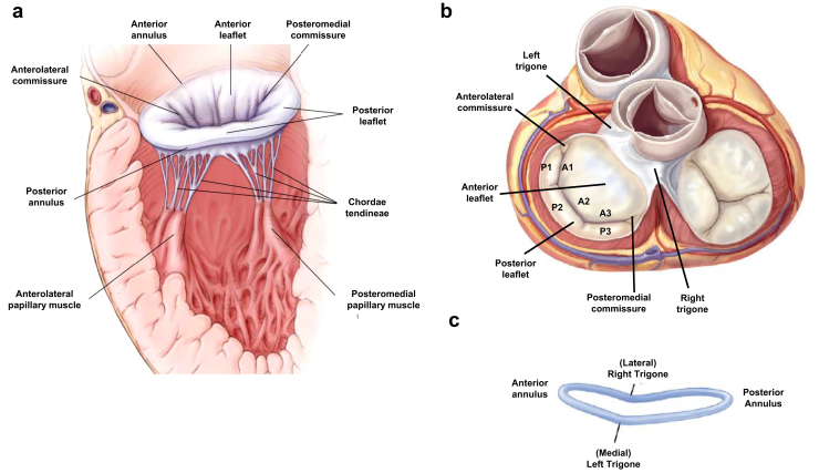
    1. McCarthy K.P., Ring L., Rana B.S. Anatomy of the mitral valve: understanding the mitral valve complex in mitral regurgitation. Eur J Echocardiogr. 2010;11:i3–i9. - PubMed
    1. Muresian H. The clinical anatomy of the mitral valve. Clin Anat. 2009;22:85–98. - PubMed
    1. Ho S.Y. Anatomy of the mitral valve. Heart. 2002;88 Suppl 4:iv5–iv10. - PMC - PubMed

LinkOut - more resources