. 2023 May 19:10:1127261.
doi: 10.3389/fcvm.2023.1127261. eCollection 2023.

Cardiomyopathy related desmocollin-2 prodomain variants affect the intracellular cadherin transport and processing

Affiliations

Cardiomyopathy related desmocollin-2 prodomain variants affect the intracellular cadherin transport and processing

Greta Marie Pohl et al. Front Cardiovasc Med. .

Abstract

Background: Arrhythmogenic cardiomyopathy can be caused by genetic variants in desmosomal cadherins. Since cardiac desmosomal cadherins are crucial for cell-cell-adhesion, their correct localization at the plasma membrane is essential.

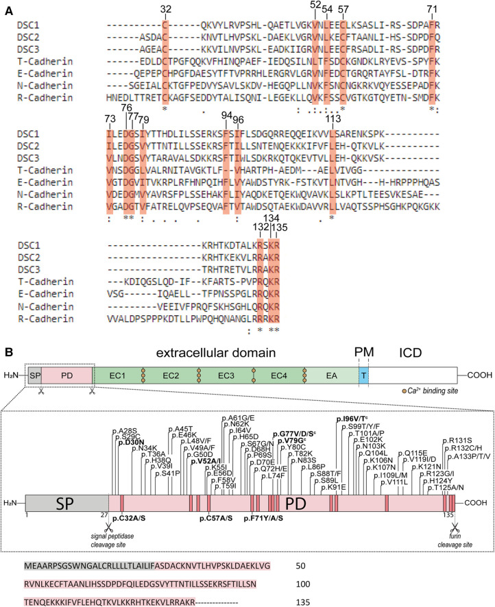
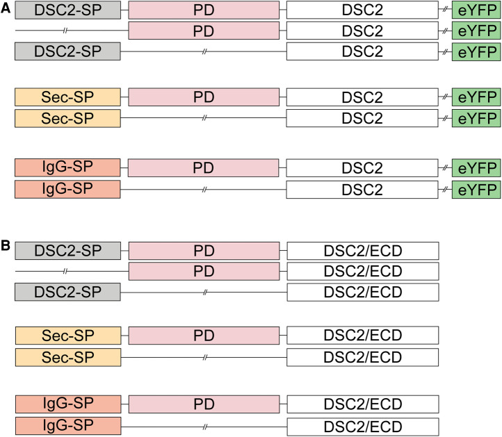
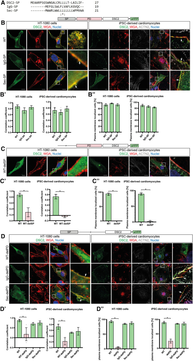
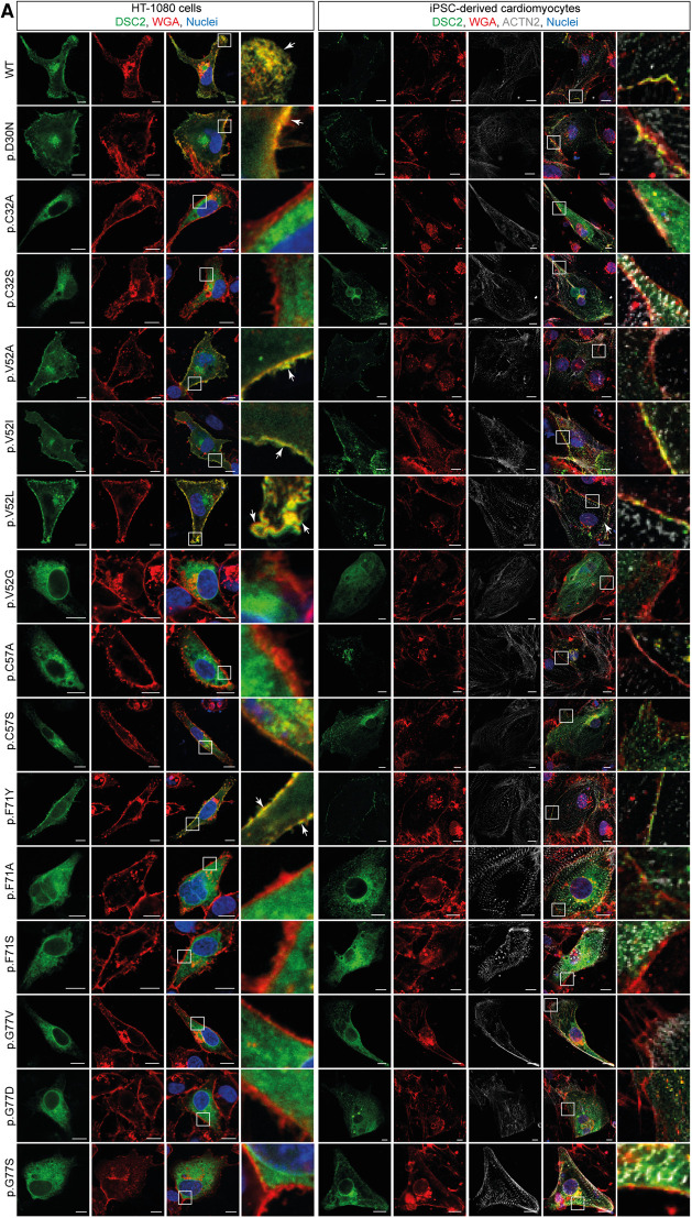
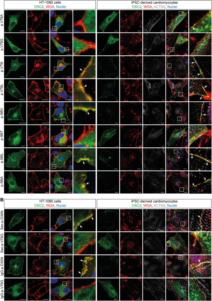
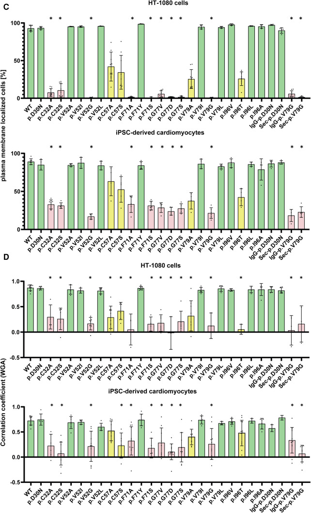
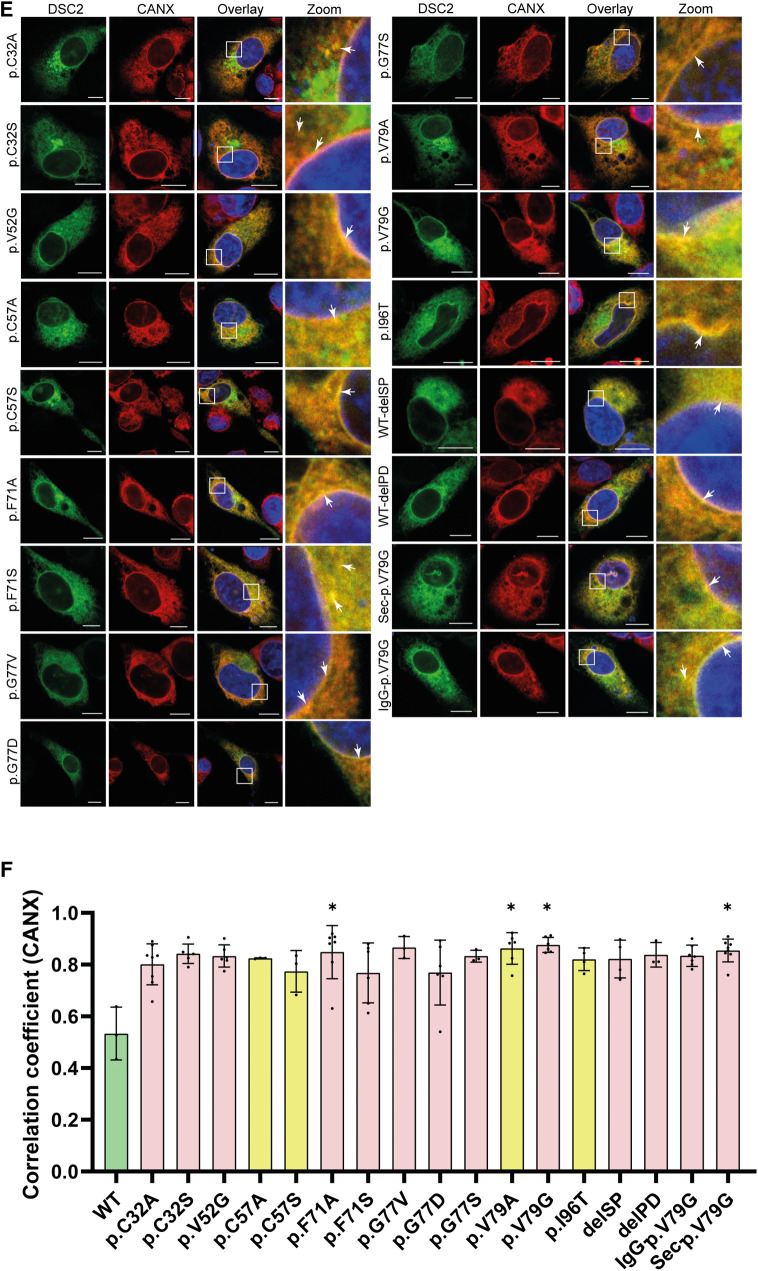
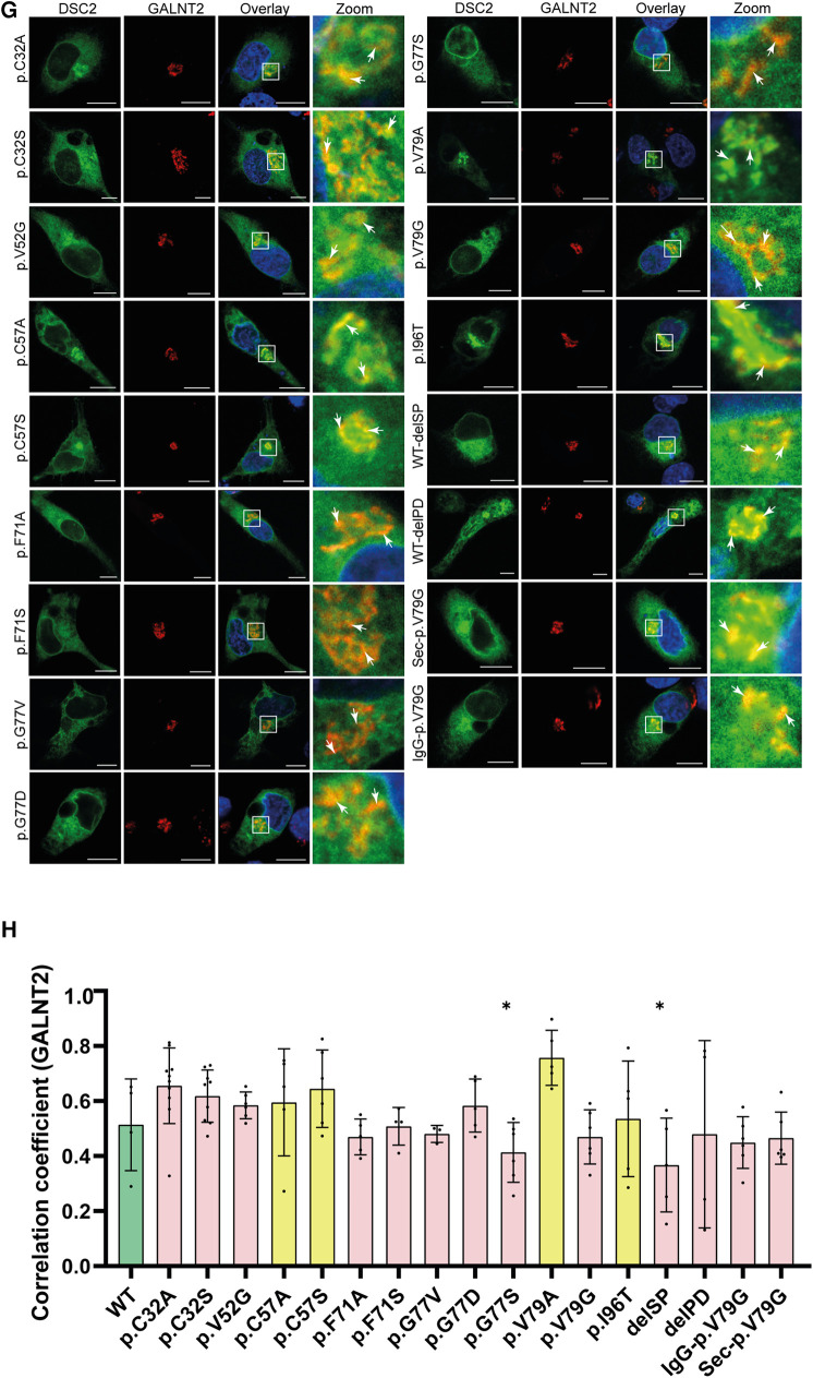
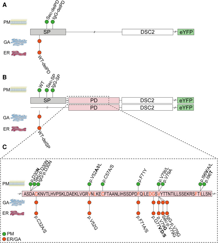
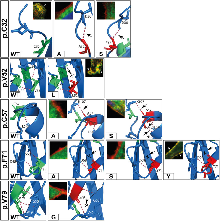
Methods: Nine desmocollin-2 variants at five positions from various public genetic databases (p.D30N, p.V52A/I, p.G77V/D/S, p.V79G, p.I96V/T) and three additional conserved positions (p.C32, p.C57, p.F71) within the prodomain were investigated in vitro using confocal microscopy. Model variants (p.C32A/S, p.V52G/L, p.C57A/S, p.F71Y/A/S, p.V79A/I/L, p.I96l/A) were generated to investigate the impact of specific amino acids.

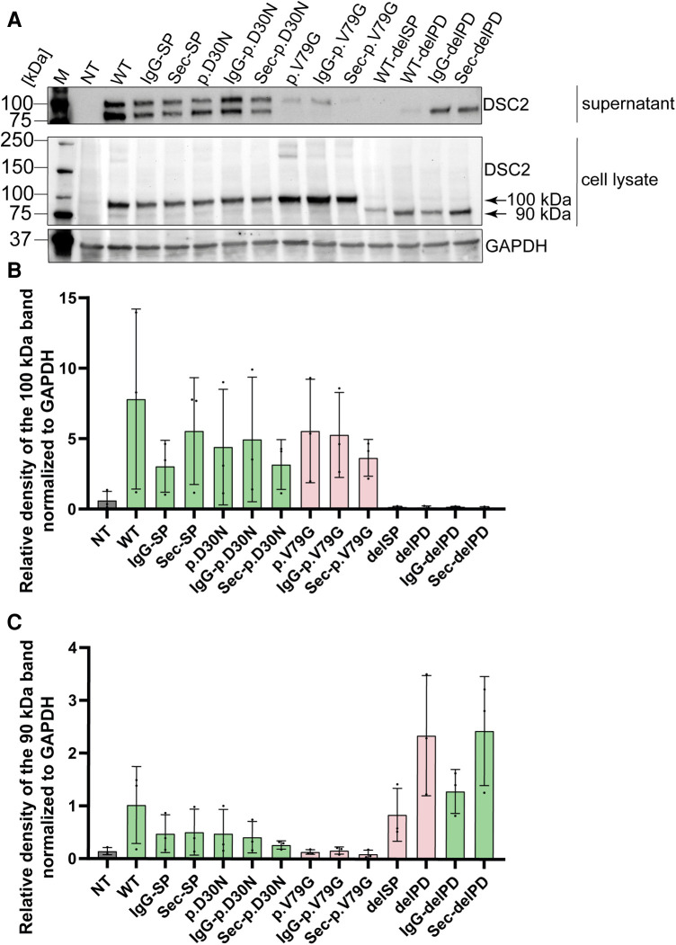
Results: We revealed that all analyzed positions in the prodomain are critical for the intracellular transport. However, the variants p.D30N, p.V52A/I and p.I96V listed in genetic databases do not disturb the intracellular transport revealing that the loss of these canonical sequences may be compensated.

Conclusion: As disease-related homozygous truncating desmocollin-2 variants lacking the transmembrane domain are not localized at the plasma membrane, we predict that some of the investigated prodomain variants may be relevant in the context of arrhythmogenic cardiomyopathy due to disturbed intracellular transport.

Keywords: DSC2; arrhythmogenic cardiomyopathy; desmocollin-2; desmosome; genetic variants; intracellular transport; prodomain; signal peptide.


Conflict of interest statement

AMS received educational grants through his institution from Abbott; Bayer Healthcare; Biosense Webster; Biotronik; Boston Scientific; BMS/Pfizer and Medtronic; and speaker/advisory board/consulting fees from Bayer Healthcare; Daiichi-Sankyo; Medtronic; Novartis and Pfizer. AB is a shareholder of Tenaya Therapeutics. The remaining authors declare that the research was conducted in the absence of any commercial or financial relationships that could be construed as a potential conflict of interest.

Figures

Figure 1
Figure 1
(A) comparison of the prodomains (PD) of DSC1-3 and different classical cadherins (N-, R-, E-Cadherin) and cadherin-like proteins (T-Cadherin) from Homo sapiens using Clustal Omega (28). Conserved amino acids are marked with orange boxes. Positions on top refer to the open reading frame (ORF) of DSC2. (B) Top: Schematic overview of the structure of DSC2. The extracellular domain consists of the signal peptide (SP, grey box), the PD (light orange box), four extracellular cadherin domains (EC1-4, green boxes) and an anchor domain (EA, light green box) which is sometimes assigned as a fifth cadherin domain (EC5). A single transmembrane domain (T, blue box) connects the extracellular region with the intracellular domain (ICD, white box). PM, plasma membrane. Middle: Schematic overview of the SP and PD of DSC2. Missense variants that are associated with ACM are shown on top and were received from the ARVC database (29), the Human Gene Mutation Database (HGMD) (30), from the National Library of Medicine ClinVar (31) and the genome aggregation database (gnomAD) (32). Variants below are investigated model mutants. All variants are classified as variants with unknown significance (VUS) according to the ACMG guidelines (27). Variants shown in bold letters were investigated. Scissors represent cleavage sites. Orange boxes mark highly conserved amino acids. c indicates variants that share conflicting interpretation of pathogenicity in various genetic databases. Bottom: Primary amino acid sequence of the SP and PD of human DSC2.
Figure 2
Figure 2
Schematic overview of the DSC2 constructs used for transient transfection of (A) HT-1080 cells and hIPSC-derived cardiomyocytes for confocal microscopy and (B) HEK293 c18 cells for the secretion assay. SP, signal peptide; PD, prodomain; eYFP, enhanced yellow fluorescence protein; DSC2/ECD, extracellular cadherin domain of desmocollin-2.
Figure 3
Figure 3
Influence of alternative signal peptides (SP) on the localization of human DSC2. (A) Amino acid alignment of the three different SPs (DSC2/IgG/Sec-SP) using Clustal Omega (28). (B) Representative images of the construct localization with different SPs, (C) DSC2 lacking the SP and (D) constructs lacking the DSC2 prodomain (PD). HT-1080 cells and hiPSC-derived cardiomyocytes were transiently transfected with different eYFP-conjugated DSC2 constructs (green). Wheat germ agglutinin (WGA) conjugated to Alexa Fluor 633 (for HT-1080) and Alexa Fluor 647 (for hiPSC-derived cardiomyocytes) was used as a plasma membrane marker and is shown in red. Anti α-Actinin (ACTN2) antibody was used as a cardiomyocyte marker (grey). Nuclei were co-stained with 4′,6-diamidino-2-phenylindole (DAPI, blue). Arrows indicate the colocalization of DSC2 and WGA. Scale bars represent 10 µm. For each construct Pearson correlation coefficient (B’, C’, D’) was determined with a region of interest of 100 µm2. At least 100 cells were manually counted blinded for plasma membrane localization by an investigator (B’’, C’’, D’’). Nonparametric Kruskal-Wallis test followed by Dunn's multiple comparison was performed using GraphPad Prism Version 9.0. P-values <0.05 were considered significant (*). Mean values ± standard deviation are shown. Of note, wildtype and alternative SP constructs are localized at the plasma membrane. Without the SP or prodomain DSC2 is not properly localized. Interestingly, with IgG- or Sec-SP but lacking the PD the protein is localized at the plasma membrane as the wildtype.
Figure 4
Figure 4
Representative images of HT-1080 cells and hiPSC-derived cardiomyocytes transiently transfected with different eYFP-conjugated DSC2 constructs. (A) Prodomain constructs in conserved positions of DSC2 (green). (B) Two representative prodomain variants (p.D30N, p.V79G) with alternative signal peptides IgG- or Sec-SP. The plasma membrane was co-stained with wheat germ agglutinin conjugated with Alexa Fluor 633 (for HT-1080) and Alexa Fluor 647 (for hiPSC-derived cardiomyocytes) (red). Anti α-Actinin (ACTN2) antibody was used as a cardiomyocyte marker (grey). (C) Statistics of DSC2 prodomain constructs localized at the plasma membrane in vitro and (D) Pearson correlation coefficient for plasma membrane localization of DSC2 prodomain constructs. Organelles were tracked using (E) anti Calnexin (CANX) antibody for co-staining of the ER and (G) anti N-acetylgalactosaminyltransferase 2 antibody (GALNT2) for the Golgi apparatus in HT-1080 cells. Nuclei were stained with 4′,6-diamidino-2-phenylindole (DAPI, blue). Scale bars represent 10 µm. Colocalization of the DSC2 prodomain constructs with the endoplasmic reticulum (ER) and the Golgi apparatus was evaluated by determining Pearson correlation coefficients with a region of interest of 100 µm2 (F,H). Green bar graphs represent variants that show plasma membrane localization, red bar graphs are variants that do not reach the membrane and yellow bar graphs represent variants that show a tendency to be poorly transported. For statistical analysis nonparametric Kruskal-Wallis test followed by Dunn's multiple comparison was used. P-values <0.05 were considered significant (*). Mean values ± standard deviation are shown. DSC2 constructs that are not transported to the plasma membrane remain within the ER or Golgi.
Figure 4
Figure 4
Representative images of HT-1080 cells and hiPSC-derived cardiomyocytes transiently transfected with different eYFP-conjugated DSC2 constructs. (A) Prodomain constructs in conserved positions of DSC2 (green). (B) Two representative prodomain variants (p.D30N, p.V79G) with alternative signal peptides IgG- or Sec-SP. The plasma membrane was co-stained with wheat germ agglutinin conjugated with Alexa Fluor 633 (for HT-1080) and Alexa Fluor 647 (for hiPSC-derived cardiomyocytes) (red). Anti α-Actinin (ACTN2) antibody was used as a cardiomyocyte marker (grey). (C) Statistics of DSC2 prodomain constructs localized at the plasma membrane in vitro and (D) Pearson correlation coefficient for plasma membrane localization of DSC2 prodomain constructs. Organelles were tracked using (E) anti Calnexin (CANX) antibody for co-staining of the ER and (G) anti N-acetylgalactosaminyltransferase 2 antibody (GALNT2) for the Golgi apparatus in HT-1080 cells. Nuclei were stained with 4′,6-diamidino-2-phenylindole (DAPI, blue). Scale bars represent 10 µm. Colocalization of the DSC2 prodomain constructs with the endoplasmic reticulum (ER) and the Golgi apparatus was evaluated by determining Pearson correlation coefficients with a region of interest of 100 µm2 (F,H). Green bar graphs represent variants that show plasma membrane localization, red bar graphs are variants that do not reach the membrane and yellow bar graphs represent variants that show a tendency to be poorly transported. For statistical analysis nonparametric Kruskal-Wallis test followed by Dunn's multiple comparison was used. P-values <0.05 were considered significant (*). Mean values ± standard deviation are shown. DSC2 constructs that are not transported to the plasma membrane remain within the ER or Golgi.
Figure 5
Figure 5
Schematic overview of the localization of different DSC2 constructs in vitro in HT-1080 cells and hiPSC-derived cardiomyocytes. (A) Different signal peptide (SP) constructs lacking the prodomain (-delPD) show localization with IgG- or Sec-SP but not with wildtype SP. (B) Different SP constructs with DSC2 prodomain (PD). Proper localization was observed when wildtype SP and IgG- or Sec-SP were conjugated to the PD. When lacking the SP (WT-delSP) DSC2 was not integrated in the plasma membrane. (C) Partial primary sequence of the prodomain of DSC2 (aa27-aa100). Conserved amino acids are given in orange. ACM-related variants are bold. ER, Endoplasmic reticulum; GA, Golgi apparatus; PM, Plasma membrane; WT, wildtype DSC2. Pictures of PM, ER and GA were taken from Servier Medical Art. Servier Medical Art by Servier is licensed under a Creative Commons Attribution 3.0 Unported License (https://creativecommons.org/licenses/by/3.0/).
Figure 6
Figure 6
Western blot analysis of secretion assay. (A) Cell culture supernatant on top and HEK293 c18 lysate (bottom). Primary antibody was used against DSC2. As loading control for the cell lysate GAPDH was used. Bar graphs showing relative density of DSC2 constructs normalized to GAPDH (B) at 100 kDa and (C) at 90 kDa. Green bar graphs represent variants which were efficiently expressed and red bar graphs show variants which were expressed but not efficiently secreted. delPD, deleted prodomain of DSC2; delSP, deleted signal peptide of DSC2; M, Marker, NT, non-transfected (negative control); WT, wildtype DSC2.
Figure 7
Figure 7
In silico predictions of polar contacts (red dashed line, arrow) in wildtype DSC2 compared to different prodomain variants. Variants that are not shown do not show different polar contacts in silico (see also Table 2). Conserved amino acids are shown in green and variants in red. Interacting amino acids are shown as sticks and are labeled. Intramolecular bonds are marked with a black arrow. The loss of a polar contact is marked with “X”. Fluorescence microscopic representative images show DSC2 prodomain variants (green) and Wheat germ agglutinin (WGA) conjugated with Alexa Fluor 633 as plasma membrane marker (red) in HT-1080 cells. Colocalization of DSC2 prodomain variants and WGA are indicated by white arrows. The variants p.V52l, p.C57A/S and p.F71Y show different intradomain contact sites but were correctly integrated in the plasma membrane. Five variants (p.C32A/S, p.F71A/S, p.V79G) were not integrated in the plasma membrane. One of the variants (p.V79G) is listed in the Human Gene Mutation Database (HGMD) and was not localized at the plasma membrane. In summary, changes of the molecular modeling structures do not correlate with the plasma membrane transport in the investiaged DSC2 constructs.

References

    1. Gumbiner BM. Regulation of cadherin-mediated adhesion in morphogenesis. Nat Rev Mol Cell Biol. (2005) 6(8):622–34. 10.1038/nrm1699 - DOI - PubMed
    1. Hulpiau P, van Roy F. Molecular evolution of the cadherin superfamily. Int J Biochem Cell Biol. (2009) 41(2):349–69. 10.1016/j.biocel.2008.09.027 - DOI - PubMed
    1. Ozawa M, Kemler R. Correct proteolytic cleavage is required for the cell adhesive function of uvomorulin. J Cell Biol. (1990) 111(4):1645–50. 10.1083/jcb.111.4.1645 - DOI - PMC - PubMed
    1. Haussinger D, Ahrens T, Aberle T, Engel J, Stetefeld J, Grzesiek S. Proteolytic E-cadherin activation followed by solution NMR and x-ray crystallography. EMBO J. (2004) 23(8):1699–708. 10.1038/sj.emboj.7600192 - DOI - PMC - PubMed
    1. Ogou SI, Yoshida-Noro C, Takeichi M. Calcium-dependent cell-cell adhesion molecules common to hepatocytes and teratocarcinoma stem cells. J Cell Biol. (1983) 97(3):944–8. 10.1083/jcb.97.3.944 - DOI - PMC - PubMed