. 2023 May 26:2023:7136819.
doi: 10.1155/2023/7136819. eCollection 2023.

Exosomal miR-141-3p from PDLSCs Alleviates High Glucose-Induced Senescence of PDLSCs by Activating the KEAP1-NRF2 Signaling Pathway

Affiliations

Exosomal miR-141-3p from PDLSCs Alleviates High Glucose-Induced Senescence of PDLSCs by Activating the KEAP1-NRF2 Signaling Pathway

Min Liu et al. Stem Cells Int. .

Abstract

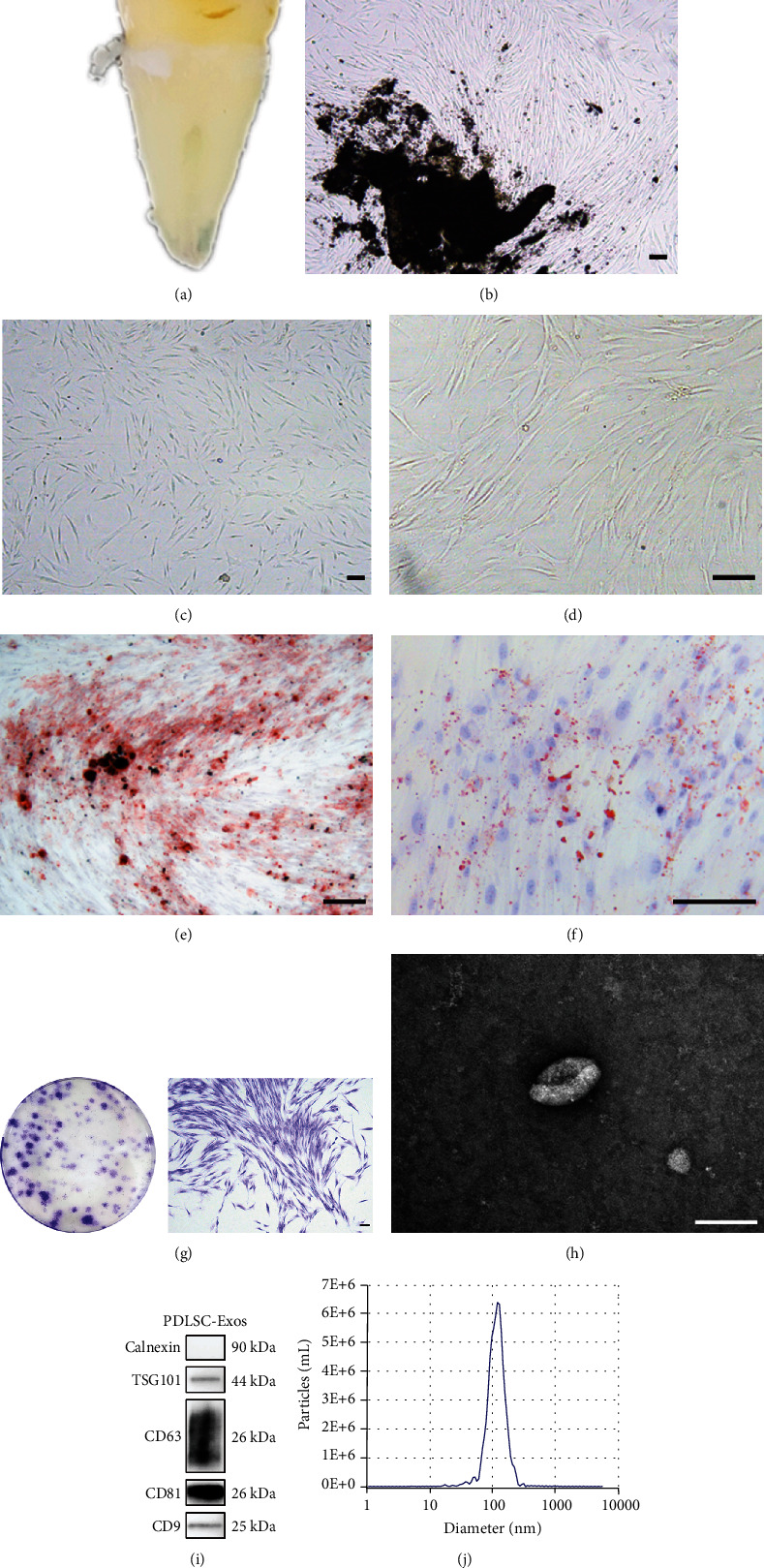
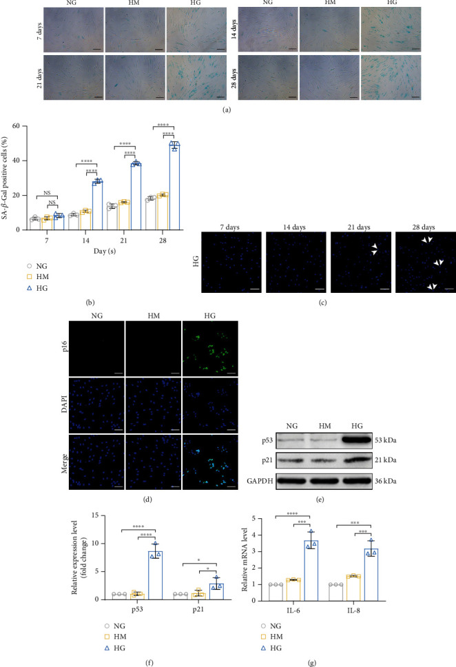
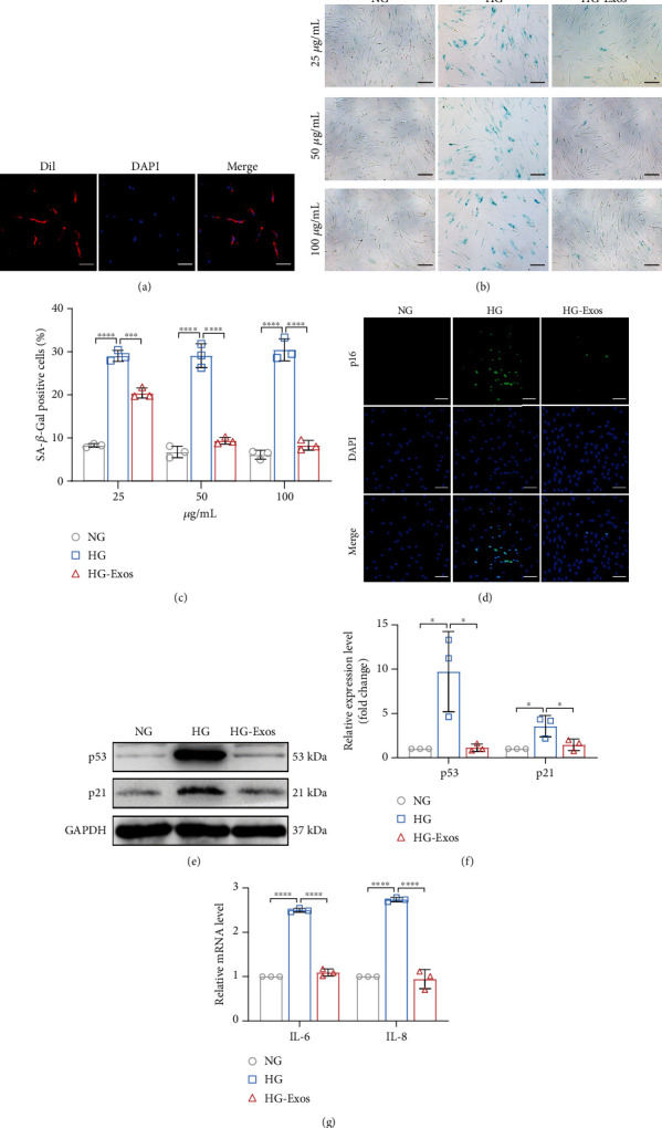
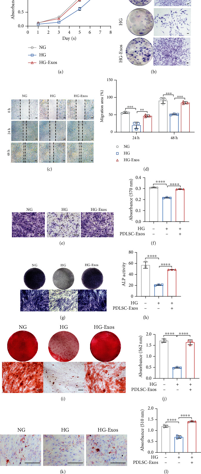
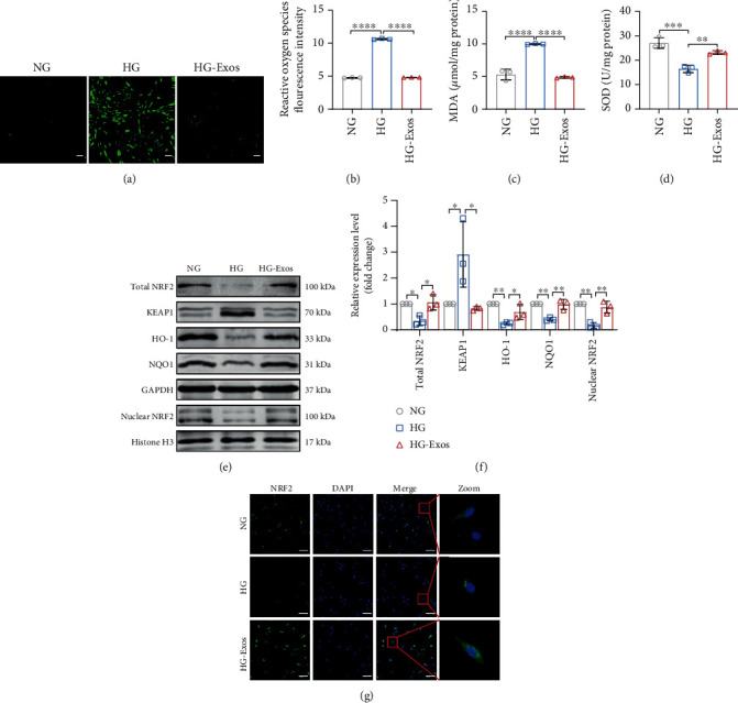
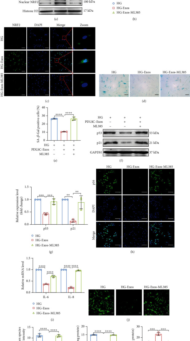
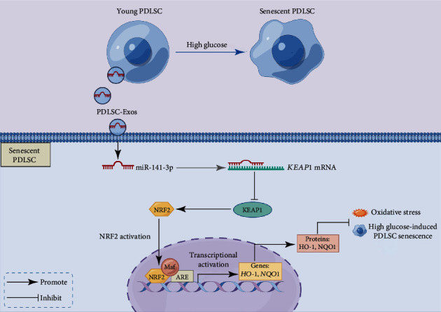
Human periodontal ligament stem cells (PDLSCs) are the most promising stem cells for periodontal tissue engineering. Senescent PDLSCs have diminished abilities to proliferate and differentiate, affecting the efficiency of periodontal tissue repair and regeneration. Stem cell-derived exosomes are important participants in intercellular information exchange and can help ameliorate senescence. In this study, we investigated PDLSC senescence in a high glucose microenvironment as well as the ability of human periodontal ligament stem cell-derived exosomes (PDLSC-Exos) to alleviate cellular senescence and the underlying mechanisms. Herein, PDLSCs and PDLSC-Exos were isolated and extracted. Then, cellular senescence indicators were evaluated after high glucose (25 mM) treatment of cultured PDLSCs. PDLSC-Exos were cocultured with senescent PDLSCs to further explore the role of PDLSC-Exos in cellular senescence and determine the differences in cellular oxidative stress levels after PDLSC-Exo treatment. Next, we investigated whether PDLSC-Exos alleviated cellular senescence by restoring the balance of oxidative stress signals and explored the underlying molecular pathways. We discovered that PDLSCs underwent premature senescence due to high glucose culture, but they were rejuvenated by PDLSC-Exos. The rejuvenating effects of PDLSC-Exos were notably reversed by cotreatment with ML385, an inhibitor of nuclear factor erythroid 2-related factor 2 (NRF2), indicating that this recovery depended on NRF2 activation. Further analyses revealed that microRNA-141-3p (miR-141-3p) was expressed at relatively high levels in PDLSC-Exos and was instrumental in PDLSC-Exo-mediated restoration by downregulating Kelch-like ECH-associated protein 1 (KEAP1), which is a negative regulator of NRF2 expression. Our findings suggest that PDLSC-Exos alleviate high glucose-induced senescence of PDLSCs by transferring miR-141-3p to activate the KEAP1-NRF2 signaling pathway. Based on this research, PDLSC-Exos may behave similarly to their parental PDLSCs and have significant effects on cellular senescence by delivering their encapsulated bioactive chemicals to target cells.


Conflict of interest statement

The authors declare that there is no conflict of interest regarding the publication of this article.

Figures

Figure 1
Figure 1
Characterization of human PDLSCs and PDLSC-Exos. (a–d) Isolation and passage of human PDLSCs. The cells exhibited a classic spindle-shaped morphology. Scale bar, 100 μm. (e) Alizarin red staining of calcified nodules indicated the capacity of PDLSCs to develop into osteoblasts. Scale bar, 100 μm. (f) Oil red O staining indicated the adipogenic differentiation capacity. Scale bar, 100 μm. (g) Clone formation assay showing the proliferation of human PDLSCs. Scale bar, 100 μm. (h) TEM observations revealed the typical cup-like shape of PDLSC-Exos. Scale bar, 100 nm. (i) WB showed that PDLSC-Exos expressed exosome marker proteins. (j) NTA revealed the particle size and concentration of PDLSC-Exos.
Figure 2
Figure 2
High glucose-induced senescence in human PDLSCs. PDLSCs were treated with normal glucose (NG group; 5.5 mM glucose), high mannitol (HM group; 5.5 mM glucose + 19.5 mM mannitol), and high glucose (HG group; 25 mM glucose) to observe phenotypic changes in senescence. (a) After 14 days of high glucose administration, an increase in the proportion of SA-β-gal-positive cells (blue) was clearly visible after SA-β-gal labeling. Scale bar, 100 μm. (b) Quantification of the percentages of SA-β-gal-positive PDLSCs in the indicated groups. (c) Cell apoptosis was observed by Hoechst 33258 staining, and apoptotic nuclei were detected as condensed (white arrows). Scale bar, 100 μm. (d) Expression of p16 (green), as measured using IF staining. Scale bar, 100 μm. (e, f) WB and quantitative analyses of cellular senescence-related protein expression (p53 and p21). (g) The expression of the indicated SASP (IL-6 and IL-8) genes was quantified using qRT–PCR. All data are presented as the means ± SDs (n = 3 independent observations). NS indicates not significant, p < 0.05, ∗∗∗p < 0.001, and ∗∗∗∗p < 0.0001. The corresponding control is indicated.
Figure 3
Figure 3
PDLSC-Exos ameliorate high glucose-induced senescence of human PDLSCs. PDLSCs were treated with high glucose levels to induce senescence and then treated with PDLSC-Exos (HG-Exos group) or PBS (HG group), while PDLSCs treated with normal glucose levels served as the control (NG group). (a) Efficient uptake of Dil-labeled exosomes by senescent PDLSCs was detected at 24 h. Scale bar, 100 μm. (b) Images of SA-β-gal-stained cells. Scale bar, 100 μm. (c) Quantification of the percentages of SA-β-gal-positive PDLSCs in the indicated groups. (d) IF detection of p16 expression (green). Scale bar, 100 μm. (e, f) The expression of p53 and p21, two senescence-related proteins, was measured and evaluated using WB. (g) The expression of the indicated SASP genes (IL-6 and IL-8) was determined using qRT–PCR. All data are presented as the means ± SDs (n = 3 independent observations). p < 0.05, ∗∗∗p < 0.001, and ∗∗∗∗p < 0.0001. The corresponding control is indicated.
Figure 4
Figure 4
PDLSC-Exos attenuate the dysfunction of high glucose-induced senescent human PDLSCs. (a, b) CCK-8 and plate colony formation assays were utilized to detect the ability of PDLSCs to proliferate. Scale bar, 100 μm. Through wound healing (c, d) and transwell experiments (e, f), cell migration was evaluated. Scale bar, 100 μm. (g, h) ALP staining/activity assays. Scale bar, 100 μm. (i, j) Alizarin red staining and quantification of mineralized nodules. Scale bar, 100 μm. (k, l) Oil red O staining and quantification of the adipogenic differentiation capacity of the cells in the three groups. Scale bar, 100 μm. All data are presented as the means ± SDs (n = 3 independent observations). ∗∗p < 0.01, ∗∗∗p < 0.001, and ∗∗∗∗p < 0.0001. The corresponding control is indicated.
Figure 5
Figure 5
PDLSC-Exos decrease oxidative stress and increase the activity of the endogenous NRF2 antioxidant system. (a, b) Intracellular ROS levels were evaluated by measuring DCFH-DA fluorescence. Scale bar, 100 μm. (c, d) The level of oxidative stress in cells was measured by detecting MDA levels and SOD activity. (e, f) Protein expression levels of total NRF2, nuclear NRF2, KEAP1, HO-1, and NQO1 in the indicated groups. (g) Localization of NRF2 (green), as visualized using IF staining. Scale bar, 100 μm. All data are presented as the means ± SDs (n = 3 independent observations). p < 0.05, ∗∗p < 0.01, ∗∗∗p < 0.001, and ∗∗∗∗p < 0.0001. The corresponding control is indicated.
Figure 6
Figure 6
PDLSC-Exos rejuvenate senescent human PDLSCs by activating NRF2. High glucose-induced senescent human PDLSCs were treated with either PDLSC-Exos alone (HG-Exos group) or in combination with an NRF2 inhibitor (HG-Exos-ML385 group), while untreated senescent PDLSCs were used as controls (HG group). (a, b) WB analysis of total NRF2, nuclear NRF2, HO-1, and NQO1 levels in the indicated groups. (c) IF staining to detect the expression and localization of NRF2 (green). Scale bar, 100 μm. (d, e) Cells were stained with SA-β-gal, and activity was determined by counting the number of SA-β-gal-positive cells. Scale bar, 100 μm. (f, g) WB and quantification of senescence-related proteins (p53 and p21). (h) IF staining indicating p16 expression. Scale bar, 100 μm. (i) SASP gene (IL-6 and IL-8) expression levels were assessed using qRT–PCR. (j, k) Cellular ROS levels were detected and quantified by performing DCFH-DA staining. Scale bar, 100 μm. (l, m) MDA levels and SOD activity were determined. All data are presented as the means ± SDs (n = 3 independent observations). p < 0.05, ∗∗p < 0.01, ∗∗∗p < 0.001, and ∗∗∗∗p < 0.0001. The corresponding control is indicated.
Figure 7
Figure 7
miR-141-3p transferred by PDLSC-Exos activates NRF2 signaling by reducing KEAP1 expression. (a) Venn diagram showing the miRNAs (hsa-miR-141-3p and hsa-miR-200a-3p) predicted to target the KEAP1 gene. (b) The expression levels of miRNAs in young and senescent PDLSCs were measured by qRT–PCR. (c) The expression levels of miRNAs in PDLSC-Exos were measured by qRT–PCR. (d) miR-141-3p expression was measured by qRT–PCR analysis. (e, f) Verification of the targeting relationship between miR-141-3p and KEAP1 using a dual-luciferase reporter gene assay. (g) qRT–PCR analysis was used to determine the expression levels of miR-141-3p in exosomes from treated human PDLSCs. NCI-Exos or 141I-Exos were used to treat high glucose-induced senescent human PDLSCs (HG-NCI-Exos group or HG-141I-Exos group), while untreated aged PDLSCs served as the control (HG group). (h, i) Total NRF2, nuclear NRF2, KEAP1, HO-1, and NQO1 protein levels were measured using WB and quantified using ImageJ software. (j) NRF2 expression levels were quantified, and NRF2 localization was identified by performing IF staining. Scale bar, 100 μm. All data are presented as the means ± SDs (n = 3 independent observations). NS indicates not significant, p < 0.05, ∗∗p < 0.01, ∗∗∗p < 0.001, and ∗∗∗∗p < 0.0001. The corresponding control is indicated.
Figure 8
Figure 8
Exosomal miR-141-3p from PDLSCs is essential for preventing PDLSC senescence caused by high-glucose treatment. (a, b) Representative image of SA-β-gal staining along with the percentage of SA-β-gal-positive cells. Scale bar, 100 μm. (c, d) The expression levels of p53 and p21 were analyzed using WB. (e) Expression of p16 detected using IF staining. Scale bar, 100 μm. (f) Relative levels of the SASP genes IL-6 and IL-8. (g, h) Cellular ROS levels were detected and quantified by DCFH-DA staining. Scale bar, 100 μm. (i, j) MDA levels and SOD activity were assessed to evaluate the level of oxidative stress in cells. All data are presented as the means ± SDs (n = 3 independent observations). p < 0.05, ∗∗p < 0.01, ∗∗∗p < 0.001, and ∗∗∗∗p < 0.0001. The corresponding control is indicated.
Figure 9
Figure 9
Schematic of the rejuvenating effects of exosomes produced by PDLSCs. High glucose levels accelerated cellular senescence in PDLSCs. PDLSC-Exos protected against high glucose-induced PDLSC senescence by transferring miR-141-3p to senescent PDLSCs to decrease KEAP1 expression and activate the NRF2 antioxidant pathway. (This image was created using Figdraw).

Similar articles

Cited by

References

    1. Lovic D., Piperidou A., Zografou I., Grassos H., Pittaras A., Manolis A. The growing epidemic of diabetes mellitus. Current Vascular Pharmacology . 2020;18(2):104–109. doi: 10.2174/1570161117666190405165911. - DOI - PubMed
    1. Genco R. J., Borgnakke W. S. Diabetes as a potential risk for periodontitis: association studies. Periodontology 2000 . 2020;83(1):40–45. doi: 10.1111/prd.12270. - DOI - PubMed
    1. Nunez J., Vignoletti F., Caffesse R. G., Sanz M. Cellular therapy in periodontal regeneration. Periodontology 2000 . 2019;79(1):107–116. doi: 10.1111/prd.12250. - DOI - PubMed
    1. Graves D. T., Ding Z., Yang Y. The impact of diabetes on periodontal diseases. Periodontology 2000 . 2020;82(1):214–224. doi: 10.1111/prd.12318. - DOI - PubMed
    1. Hayelick L. The limited in vitro lifetime of human diploid cell strains. Experimental Cell Research . 1965;37(3):614–636. doi: 10.1016/0014-4827(65)90211-9. - DOI - PubMed