Modeling of active skeletal muscles: a 3D continuum approach incorporating multiple muscle interactions
- PMID: 37274172
- PMCID: PMC10234509
- DOI: 10.3389/fbioe.2023.1153692
Modeling of active skeletal muscles: a 3D continuum approach incorporating multiple muscle interactions
Abstract
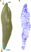
Skeletal muscles have a highly organized hierarchical structure, whose main function is to generate forces for movement and stability. To understand the complex heterogeneous behaviors of muscles, computational modeling has advanced as a non-invasive approach to evaluate relevant mechanical quantities. Aiming to improve musculoskeletal predictions, this paper presents a framework for modeling 3D deformable muscles that includes continuum constitutive representation, parametric determination, model validation, fiber distribution estimation, and integration of multiple muscles into a system level for joint motion simulation. The passive and active muscle properties were modeled based on the strain energy approach with Hill-type hyperelastic constitutive laws. A parametric study was conducted to validate the model using experimental datasets of passive and active rabbit leg muscles. The active muscle model with calibrated material parameters was then implemented to simulate knee bending during a squat with multiple quadriceps muscles. A computational fluid dynamics (CFD) fiber simulation approach was utilized to estimate the fiber arrangements for each muscle, and a cohesive contact approach was applied to simulate the interactions among muscles. The single muscle simulation results showed that both passive and active muscle elongation responses matched the range of the testing data. The dynamic simulation of knee flexion and extension showed the predictive capability of the model for estimating the active quadriceps responses, which indicates that the presented modeling pipeline is effective and stable for simulating multiple muscle configurations. This work provided an effective framework of a 3D continuum muscle model for complex muscle behavior simulation, which will facilitate additional computational and experimental studies of skeletal muscle mechanics. This study will offer valuable insight into the future development of multiscale neuromuscular models and applications of these models to a wide variety of relevant areas such as biomechanics and clinical research.
Keywords: Hill-type muscle model; active skeletal muscle; finite element analysis; muscle interactions; parametric study; quadriceps.
Copyright © 2023 Zeng, Hume, Lu, Fitzpatrick, Babcock, Myers, Rullkoetter and Shelburne.
Conflict of interest statement
The authors declare that the research was conducted in the absence of any commercial or financial relationships that could be construed as a potential conflict of interest.
Figures


Similar articles
-
Development of a finite element musculoskeletal model with the ability to predict contractions of three-dimensional muscles.J Biomech. 2019 Sep 20;94:230-234. doi: 10.1016/j.jbiomech.2019.07.042. Epub 2019 Aug 7. J Biomech. 2019. PMID: 31421809
-
Constitutive Models for Active Skeletal Muscle: Review, Comparison, and Application in a Novel Continuum Shoulder Model.Int J Numer Method Biomed Eng. 2025 Apr;41(4):e70036. doi: 10.1002/cnm.70036. Int J Numer Method Biomed Eng. 2025. PMID: 40267885 Free PMC article. Review.
-
A finite-element model for the mechanical analysis of skeletal muscles.J Theor Biol. 2000 Sep 7;206(1):131-49. doi: 10.1006/jtbi.2000.2109. J Theor Biol. 2000. PMID: 10968943
-
A computationally efficient strategy to estimate muscle forces in a finite element musculoskeletal model of the lower limb.J Biomech. 2019 Feb 14;84:94-102. doi: 10.1016/j.jbiomech.2018.12.020. Epub 2018 Dec 28. J Biomech. 2019. PMID: 30616983 Free PMC article.
-
A Systematic Review of Continuum Modeling of Skeletal Muscles: Current Trends, Limitations, and Recommendations.Appl Bionics Biomech. 2018 Dec 6;2018:7631818. doi: 10.1155/2018/7631818. eCollection 2018. Appl Bionics Biomech. 2018. PMID: 30627216 Free PMC article. Review.
Cited by
-
A Biomechanical Simulation of Forearm Flexion Using the Finite Element Approach.Bioengineering (Basel). 2023 Dec 25;11(1):23. doi: 10.3390/bioengineering11010023. Bioengineering (Basel). 2023. PMID: 38247900 Free PMC article.
-
Emerging Innovations in Preoperative Planning and Motion Analysis in Orthopedic Surgery.Diagnostics (Basel). 2024 Jun 21;14(13):1321. doi: 10.3390/diagnostics14131321. Diagnostics (Basel). 2024. PMID: 39001212 Free PMC article. Review.
-
Neural-driven activation of 3D muscle within a finite element framework: exploring applications in healthy and neurodegenerative simulations.Comput Methods Biomech Biomed Engin. 2024 Dec;27(16):2389-2399. doi: 10.1080/10255842.2023.2280772. Epub 2023 Nov 15. Comput Methods Biomech Biomed Engin. 2024. PMID: 37966863
-
Neuromusculoskeletal Modeling and Force Prediction: Verification Through Experimental Neuromuscular Dynamics.Ann Biomed Eng. 2025 Jul 8. doi: 10.1007/s10439-025-03783-2. Online ahead of print. Ann Biomed Eng. 2025. PMID: 40627083
-
Determining a musculoskeletal system's pre-stretched state using continuum-mechanical forward modelling and joint range optimization.Biomech Model Mechanobiol. 2024 Jun;23(3):1031-1053. doi: 10.1007/s10237-024-01821-x. Epub 2024 Apr 15. Biomech Model Mechanobiol. 2024. PMID: 38619712 Free PMC article.
References
-
- Ali A. A., Shalhoub S. S., Cyr A. J., Fitzpatrick C. K., Maletsky L. P., Rullkoetter P. J., et al. (2016). Validation of predicted patellofemoral mechanics in a finite element model of the healthy and cruciate-deficient knee. J. Biomech. 49 (2), 302–309. 10.1016/j.jbiomech.2015.12.020 - DOI - PMC - PubMed
Grants and funding
LinkOut - more resources
Full Text Sources
Miscellaneous

