

Highlighting the novel effects of high-intensity interval training on some histopathological and molecular indices in the heart of type 2 diabetic rats
- PMID: 37274326
- PMCID: PMC10235768
- DOI: 10.3389/fendo.2023.1175585
Highlighting the novel effects of high-intensity interval training on some histopathological and molecular indices in the heart of type 2 diabetic rats
Abstract
Background: Type 2 diabetes is one of the most common metabolic diseases in recent years and has become an important risk factor for cardiovascular disorders. The first goal is to reduce type 2 diabetes, and in the case of cardiovascular disease, the second goal is to reduce and manage that disorder.
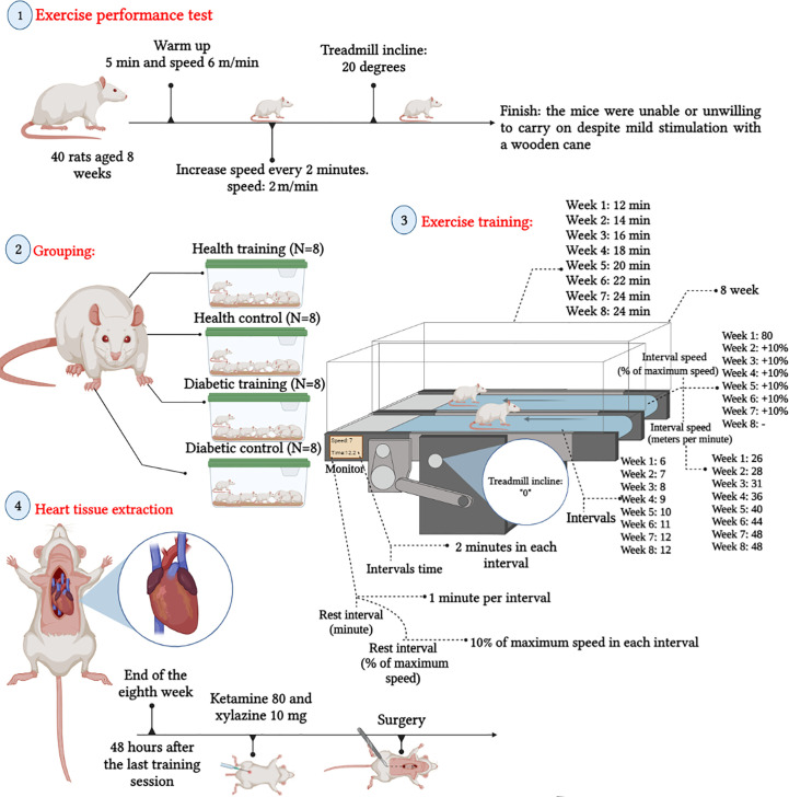
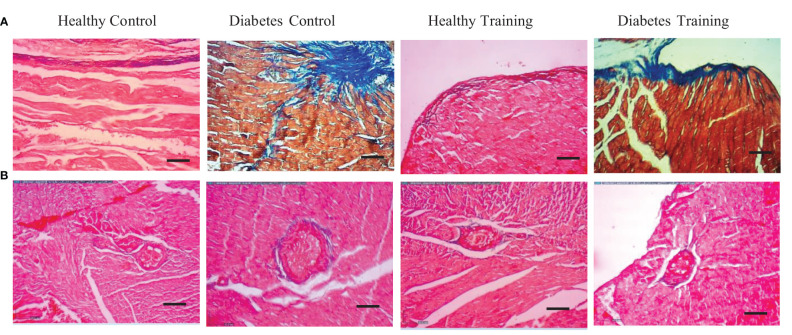
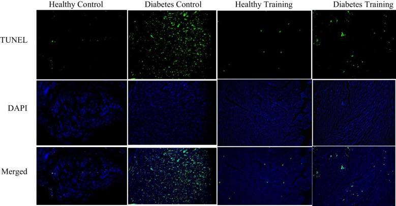
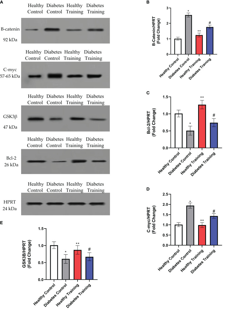
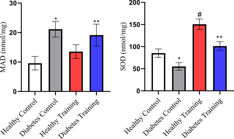
Materials and methods: The rats were divided into 4 groups: Healthy Control (n=8), Diabetes Control (n=8), Diabetes Training (n=8), and Healthy Training (n=8). The protocol consisted of 8 weeks of High-intensity interval (5 sessions per week), where the training started with 80% of the peak speed in the first week, and 10% was added to this speed every week. To measure the level of B-catenin, c-MYC, GSK3B, and Bcl-2 proteins using the western blot method, cardiac pathological changes were measured using hematoxylin and eosin staining, Masson's trichrome and PAS staining and apoptosis using the TUNEL method.
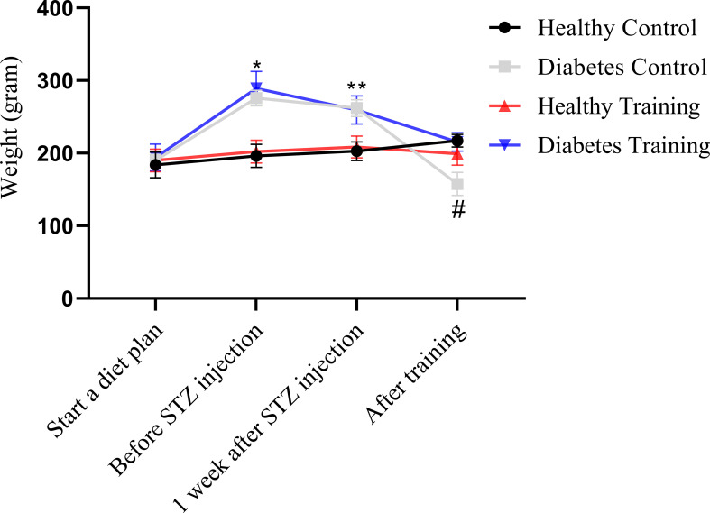
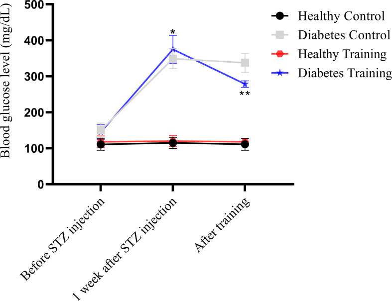
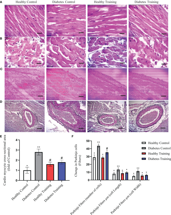
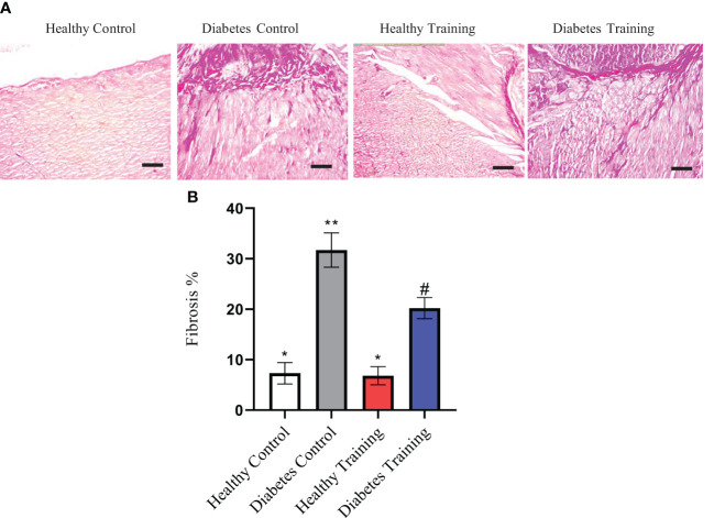
Findings: Histological results showed that diabetes causes significant pathological hypertrophy, fibrosis, and severe apoptosis in heart tissue. HIIT training significantly reduced pathological hypertrophy and fibrosis in heart tissue, and the rate of cardiomyocyte apoptosis was greatly reduced. This research showed that diabetes disorder increases the levels of B-catenin and c-Myc proteins and causes a decrease in the expression of GSK3B and Bcl-2 proteins. After eight weeks of HIIT training, the levels of B-catenin and c-Myc proteins decreased significantly, and the levels of GSK3B and Bcl-2 proteins increased.
Conclusion: This study showed that HIIT could be a suitable strategy to reduce cardiomyopathy in type 2 diabetic rats. However, it is suggested that in future studies, researchers should perform different intensities and exercises to promote exercise goals in type 2 diabetic cardiomyopathy.
Keywords: cardiomyopathy; diabetic; exercise; high-intensity interval training; physical activity.
Copyright © 2023 Rami, Rahdar, Ahmadi Hekmatikar and Awang Daud.
Conflict of interest statement
The authors declare that the research was conducted in the absence of any commercial or financial relationships that could be construed as a potential conflict of interest.
Figures
Similar articles
-
Highlighting the effects of high-intensity interval training on the changes associated with hypertrophy, apoptosis, and histological proteins of the heart of old rats with type 2 diabetes.Sci Rep. 2024 Mar 26;14(1):7133. doi: 10.1038/s41598-024-57119-6. Sci Rep. 2024. PMID: 38531890 Free PMC article.
-
Lung molecular and histological changes in type 2 diabetic rats and its improvement by high-intensity interval training.BMC Pulm Med. 2024 Jan 17;24(1):37. doi: 10.1186/s12890-024-02840-1. BMC Pulm Med. 2024. PMID: 38233819 Free PMC article.
-
The effect of 8 weeks of high-intensity interval training and moderate-intensity continuous training on cardiac angiogenesis factor in diabetic male rats.J Physiol Biochem. 2020 May;76(2):291-299. doi: 10.1007/s13105-020-00733-5. Epub 2020 Mar 10. J Physiol Biochem. 2020. PMID: 32157499
-
STAT4 regulates cardiomyocyte apoptosis in rat models of diabetic cardiomyopathy.Acta Histochem. 2022 May;124(4):151872. doi: 10.1016/j.acthis.2022.151872. Epub 2022 Mar 31. Acta Histochem. 2022. PMID: 35367814
-
Effects of High-Intensity Interval Training on People Living with Type 2 Diabetes: A Narrative Review.Can J Diabetes. 2017 Oct;41(5):536-547. doi: 10.1016/j.jcjd.2016.12.004. Epub 2017 Mar 30. Can J Diabetes. 2017. PMID: 28366674 Review.
Cited by
-
Combined Aerobic Exercise with Intermittent Fasting Is Effective for Reducing mTOR and Bcl-2 Levels in Obese Females.Sports (Basel). 2024 Apr 25;12(5):116. doi: 10.3390/sports12050116. Sports (Basel). 2024. PMID: 38786985 Free PMC article.
-
Exercise and dietary interventions in the management of diabetic cardiomyopathy: mechanisms and implications.Cardiovasc Diabetol. 2025 Apr 9;24(1):159. doi: 10.1186/s12933-025-02702-y. Cardiovasc Diabetol. 2025. PMID: 40205621 Free PMC article. Review.
-
Protective effects of aerobic exercise on cardiac histology and stereological parameters in a rat model of type 2 diabetes mellitus.J Diabetes Metab Disord. 2025 Jun 2;24(1):138. doi: 10.1007/s40200-025-01641-5. eCollection 2025 Jun. J Diabetes Metab Disord. 2025. PMID: 40469910
-
Highlighting the effects of high-intensity interval training on the changes associated with hypertrophy, apoptosis, and histological proteins of the heart of old rats with type 2 diabetes.Sci Rep. 2024 Mar 26;14(1):7133. doi: 10.1038/s41598-024-57119-6. Sci Rep. 2024. PMID: 38531890 Free PMC article.
-
Narrative Review of High-Intensity Interval Training: Positive Impacts on Cardiovascular Health and Disease Prevention.J Cardiovasc Dev Dis. 2025 Apr 17;12(4):158. doi: 10.3390/jcdd12040158. J Cardiovasc Dev Dis. 2025. PMID: 40278218 Free PMC article. Review.
References
-
- Kurdiova T, Balaz M, Vician M, Maderova D, Vlcek M, Valkovic L, et al. . Effects of obesity, diabetes and exercise on Fndc5 gene expression and irisin release in human skeletal muscle and adipose tissue: in vivo and in vitro studies. J Physiol (2014) 592(5):1091–107. doi: 10.1113/jphysiol.2013.264655 - DOI - PMC - PubMed
Publication types
MeSH terms
Substances
LinkOut - more resources
Full Text Sources
Medical
Miscellaneous