

Rituximab exerts its anti-arthritic effects via inhibiting NF-κB/GM-CSF/iNOS signaling in B cells in a mouse model of collagen-induced arthritis
- PMID: 37274671
- PMCID: PMC10238934
- DOI: 10.1016/j.heliyon.2023.e16673
Rituximab exerts its anti-arthritic effects via inhibiting NF-κB/GM-CSF/iNOS signaling in B cells in a mouse model of collagen-induced arthritis
Abstract
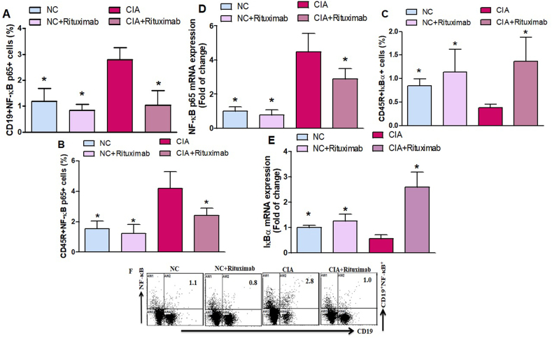
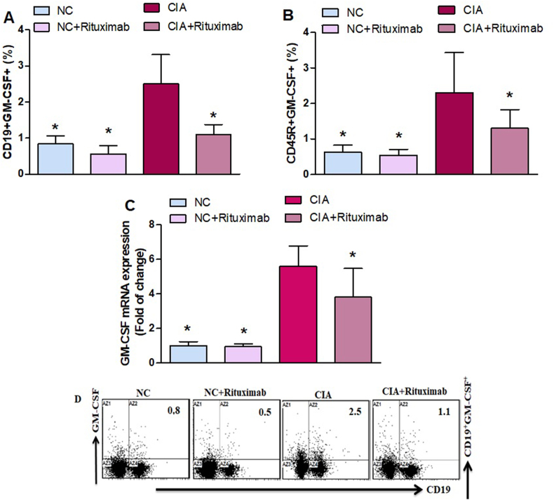
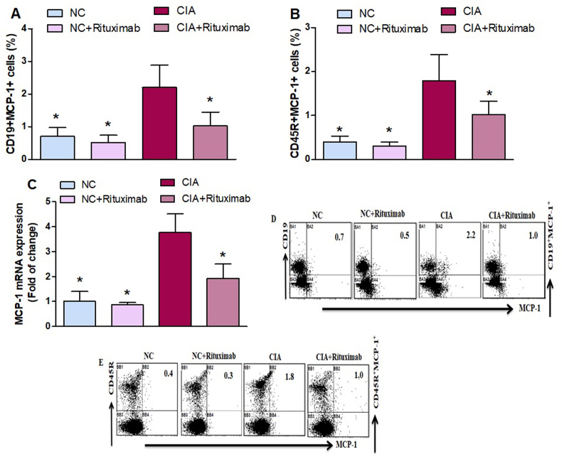
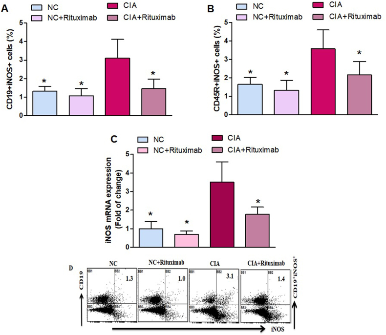
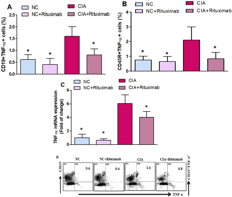
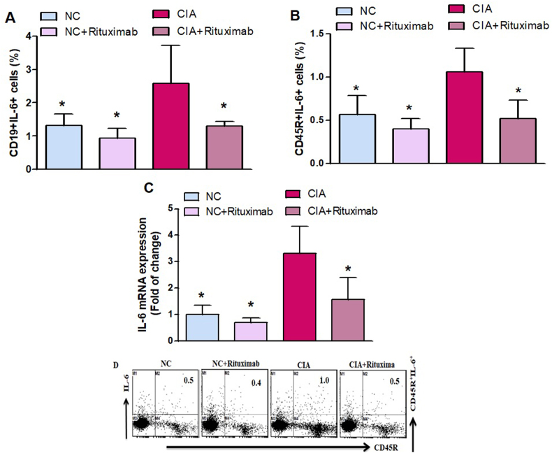
Rheumatoidarthritis (RA) is an autoimmune disease characterized by uncontrolled joint inflammation and damage to bone and cartilage. B cells are known to play a crucial role in the pathogenesis and development of arthritis. Previous studies have found that B cells may be a potential target for treating RA. Rituximab, a monoclonal antibody targeting B cells, has induced long-term clinical responses in RA. Collagen-induced arthritis (CIA) mouse model is a widely studied autoimmune model of RA. CIA mouse model was used to investigate the effect of rituximab on the RA severity in the mice. Following induction of CIA, animals were treated with rituximab (250 mg/kg/week) intraperitoneally on the days 28, 35, 42, 49, 56, and 63 after collagen induction. We investigated the effect of rituximab on NF-κB p65, IκBα, GM-CSF, MCP-1, iNOS, TNF-α, and IL-6 cells in splenic CD19+ and CD45R+ B cells using flow cytometry. We also assessed the effect of rituximab on NF-κB p65, GM-CSF, IκBα, MCP-1, iNOS, TNF-α, and IL-6 at mRNA levels using RT-PCR analyses of knee tissues. Rituximab treatment significantly decreased CD19+NF-κB p65+, CD45R+NF-κB p65+, CD19+GM-CSF+, CD45R+GM-CSF+, CD19+MCP-1+, CD45R+MCP-1+, CD19+TNF-α+, CD45R+TNF-α+, CD19+iNOS+, CD45R+iNOS+, CD19+IL-6+, and CD45R+IL-6+, and increased CD45R+IκBα+ in spleen cells of CIA mice. We further observed that rituximab treatment downregulated NF-κB p65, GM-CSF, MCP-1, iNOS, TNF-α, and IL-6, whereas it upregulated IκBα, mRNA level. All these findings suggest that rituximab may be a novel therapeutic target for the treatment of RA.
Keywords: Autoimmune disease; B cells; Collagen-induced arthritis; NF-κB/GM-CSF/iNOS signaling; Rituximab.
© 2023 The Authors.
Conflict of interest statement
The authors declare that they have no known competing financial interests or personal relationships that could have appeared to influence the work reported in this paper.
Figures
Similar articles
-
Mitogen-activated protein kinase inhibitor PD98059 improves neuroimmune dysfunction in experimental autoimmune encephalomyelitis in SJL/J mice through the inhibition of nuclear factor-kappa B signaling in B cells.Brain Res Bull. 2023 Mar;194:45-53. doi: 10.1016/j.brainresbull.2023.01.003. Epub 2023 Jan 13. Brain Res Bull. 2023. PMID: 36646144
-
Cadmium exposure exacerbates immunological abnormalities in a BTBR T+ Itpr3tf/J autistic mouse model by upregulating inflammatory mediators in CD45R-expressing cells.J Neuroimmunol. 2024 Jan 15;386:578253. doi: 10.1016/j.jneuroim.2023.578253. Epub 2023 Dec 3. J Neuroimmunol. 2024. PMID: 38064869
-
CXC chemokine receptor 3 antagonist AMG487 shows potent anti-arthritic effects on collagen-induced arthritis by modifying B cell inflammatory profile.Immunol Lett. 2020 Sep;225:74-81. doi: 10.1016/j.imlet.2020.06.014. Epub 2020 Jun 23. Immunol Lett. 2020. PMID: 32590119
-
DAPTA, a C-C Chemokine Receptor 5 (CCR5), Leads to the Downregulation of Notch/NF-κB Signaling and Proinflammatory Mediators in CD40+ Cells in Experimental Autoimmune Encephalomyelitis Model in SJL/J Mice.Biomedicines. 2023 May 23;11(6):1511. doi: 10.3390/biomedicines11061511. Biomedicines. 2023. PMID: 37371605 Free PMC article.
-
Histamine H4 Receptor Agonist, 4-Methylhistamine, Aggravates Disease Progression and Promotes Pro-Inflammatory Signaling in B Cells in an Experimental Autoimmune Encephalomyelitis Mouse Model.Int J Mol Sci. 2023 Aug 20;24(16):12991. doi: 10.3390/ijms241612991. Int J Mol Sci. 2023. PMID: 37629172 Free PMC article.
Cited by
-
Nonspecific Orbital Inflammation (NSOI): Unraveling the Molecular Pathogenesis, Diagnostic Modalities, and Therapeutic Interventions.Int J Mol Sci. 2024 Jan 26;25(3):1553. doi: 10.3390/ijms25031553. Int J Mol Sci. 2024. PMID: 38338832 Free PMC article. Review.
-
Exploring the causal relationship between interleukin levels and osteoarthritis: A two-sample Mendelian randomization analysis.Medicine (Baltimore). 2025 Jun 6;104(23):e42675. doi: 10.1097/MD.0000000000042675. Medicine (Baltimore). 2025. PMID: 40489884 Free PMC article.
-
Potential Rheumatoid Arthritis-Associated Interstitial Lung Disease Treatment and Computational Approach for Future Drug Development.Int J Mol Sci. 2024 Feb 26;25(5):2682. doi: 10.3390/ijms25052682. Int J Mol Sci. 2024. PMID: 38473928 Free PMC article. Review.
References
LinkOut - more resources
Full Text Sources
Research Materials
Miscellaneous
