

Rathke Cleft Cyst with Occulomotor Palsy: An Unusual Presentation
- PMID: 37275054
- PMCID: PMC10235355
- DOI: 10.1007/s12070-022-03283-y
Rathke Cleft Cyst with Occulomotor Palsy: An Unusual Presentation
Abstract
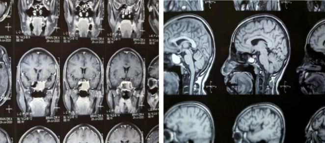
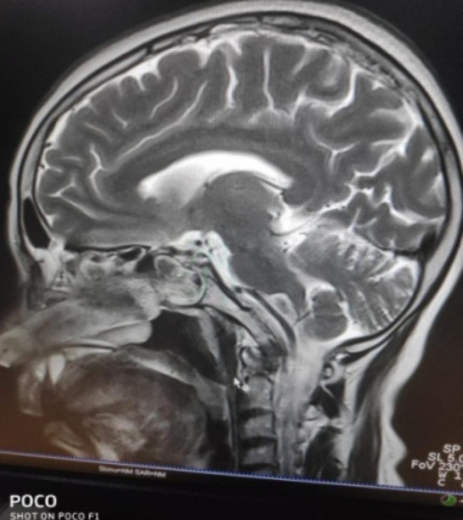
Rathke cleft cysts arise from remnants of Rathke's pouch. Often identified as incidental autopsy findings, these cysts can occasionally become large enough to cause headache, vision impairment, hypothalamic pituitary dysfunction and rarely, cranial neuropathy. MRI is the imaging modality of choice with surgical drainage recommended in symptomatic cases.
Keywords: Occulomotor; Palsy; Rathke cyst.
© Association of Otolaryngologists of India 2022. Springer Nature or its licensor (e.g. a society or other partner) holds exclusive rights to this article under a publishing agreement with the author(s) or other rightsholder(s); author self-archiving of the accepted manuscript version of this article is solely governed by the terms of such publishing agreement and applicable law.
Conflict of interest statement
Conflict of InterestsNil declared.
Figures


References
-
- McGrath P Cysts of sellar and pharyngeal hypophyses. Pathology. 1971 Jan 1;3(2):123 – 31 - PubMed
-
- Burger PC, Scheithauer BW (1994) Tumors of the central nervous system. Amer Registry of Pathology
-
- Trifanescu R, Ansorge O, Wass JA, Grossman AB, Karavitaki N (2012 Feb) Rathke’s cleft cysts. Clin Endocrinol 76(2):151–160 - PubMed
-
- Yum HR, Jang J, Shin SY, Park SH (2015) Rathke cleft cyst presenting as unilateral progressive oculomotor nerve palsy. Canadian Journal of Ophthalmology. Apr 1;50(2):e31-3 - PubMed
-
- Zada G (2011) Rathke cleft cysts: a review of clinical and surgical management. Neurosurgical focus. Jul 1;31(1):E1 - PubMed
LinkOut - more resources
Full Text Sources