

A Rare Case of Occipital Subdural Abscess of Rhinogenic Origin: Case Report
- PMID: 37275101
- PMCID: PMC10235282
- DOI: 10.1007/s12070-022-03131-z
A Rare Case of Occipital Subdural Abscess of Rhinogenic Origin: Case Report
Abstract
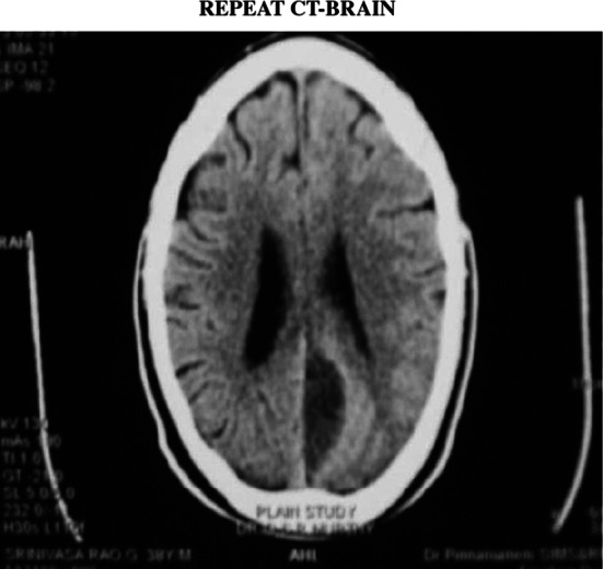
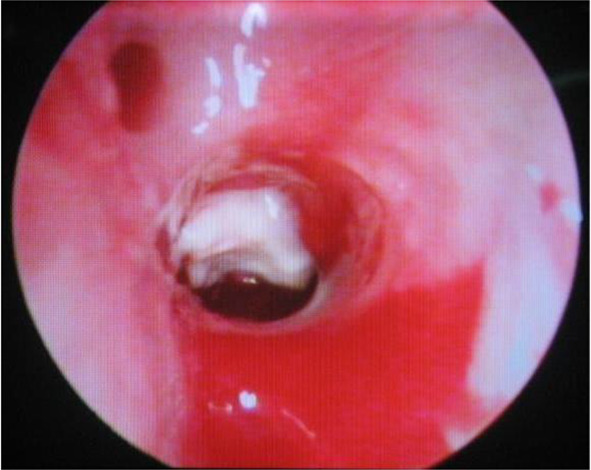
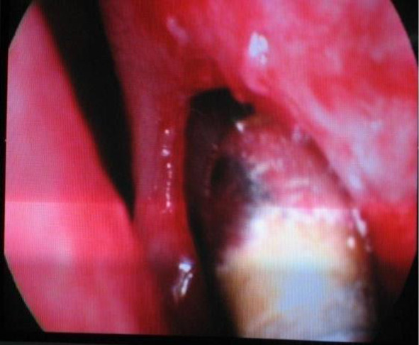
In the present day scenario complications arising from sinusitis are rare, since the introduction of antibiotics. However Sinusitis and its complications are still existing and masking of symptoms due to inadequate dosage and duration of antibiotics, which is life threatening and if neglected may result in high morbidity and mortality. This case has been presented for its rare presentation of parafalcine abscess of left occipital lobe of middle cranial fossa, which is secondary to frontal sinusitis. A 40 year old male patient was admitted in ICU with chief complaints of fever for 3 days, headache and neck stiffness for 2 days and altered sensorium for 1 day. Computed tomography showed both frontals, ethmoids and maxillary sinusitis on the left side. Patient was given conservative treatment and showed partial improvement of symptoms. On repeat CT brain (after 2 weeks) showed abscess parasagittally in left occipital lobe. Neurosurgeon aspirated abscess through parasagittal approach. Later the patient referred to the ENT department and ESS (endoscopic sinus surgery) DRAF II procedure done for clearance of frontal sinus to avoid recurrence of intracranial complications.
Keywords: DRAF II; Frontal sinusitis; Occipital lobe; Parasagittal abscess.
© Association of Otolaryngologists of India 2022. Springer Nature or its licensor holds exclusive rights to this article under a publishing agreement with the author(s) or other rightsholder(s); author self-archiving of the accepted manuscript version of this article is solely governed by the terms of such publishing agreement and applicable law.
Conflict of interest statement
Conflict of interestAll the authors declare that they have no conflict of interest.
Figures


References
-
- Som PMC, Curtin HD. Head and neck imaging. St. Louis: Mosby; 2011.
LinkOut - more resources
Full Text Sources
Research Materials
Miscellaneous