. 2023 Mar 28;7(3):100133.
doi: 10.1016/j.rpth.2023.100133. eCollection 2023 Mar.

Evaluation of four commercial ELISAs to measure tissue factor in human plasma

Affiliations

Evaluation of four commercial ELISAs to measure tissue factor in human plasma

Ana T A Sachetto et al. Res Pract Thromb Haemost. .

Abstract

Background: Under pathological conditions, tissue factor (TF)-positive extracellular vesicles (EVs) are released into the circulation and activate coagulation. Therefore, it is important to identify methods that accurately quantitate levels of TF in plasma. Enzyme-linked immunosorbent assays (ELISAs) are a fast and simple method to quantitate levels of proteins. However, there are several specific challenges with measuring TF antigen in plasma including its low concentration and the complexity of plasma.

Objectives: We aimed to evaluate the ability of 4 commercial ELISAs to measure TF in human plasma.

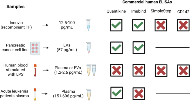
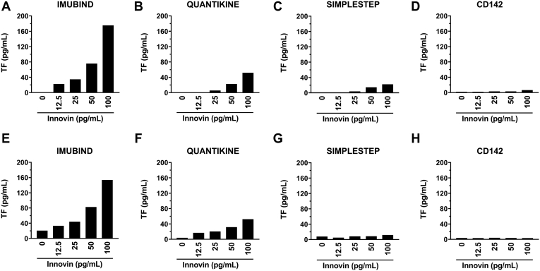
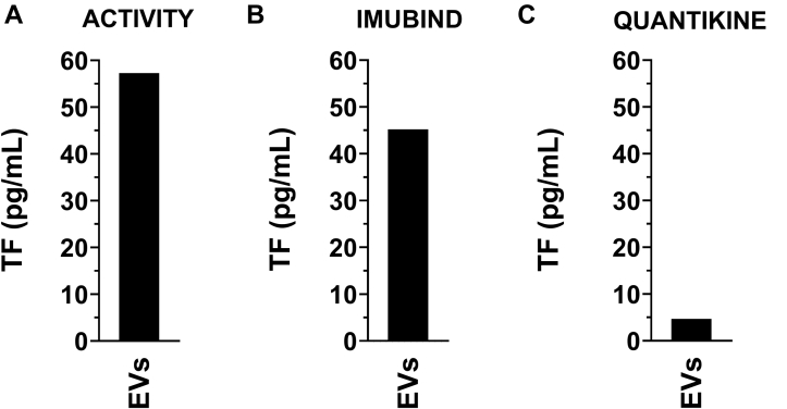
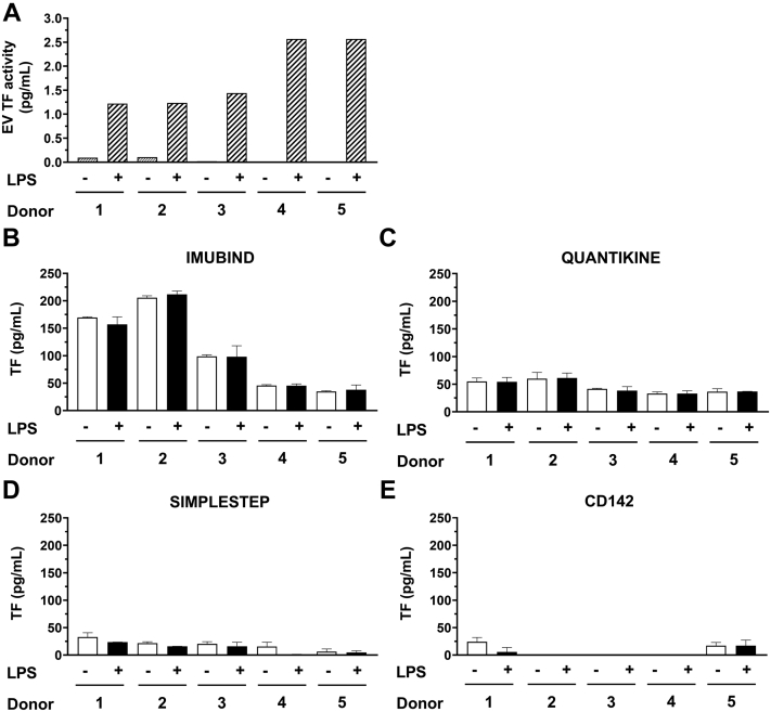
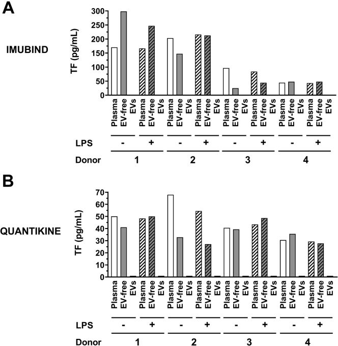
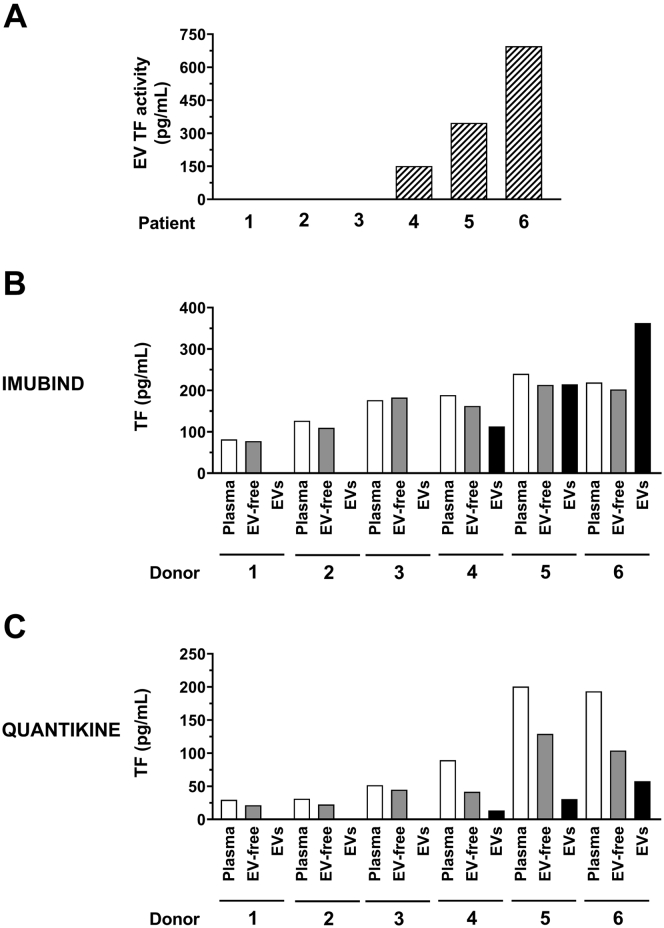
Methods: We determined the ability of 4 commercial ELISAs (Imubind, Quantikine, Human SimpleStep, and CD142 Human) to detect recombinant human TF (Innovin) (12.5-100 pg/mL), TF-positive EVs isolated from the culture supernatant from a human pancreatic cancer cell line (57 pg/mL), TF in plasma containing low levels of EV TF activity (1.2-2.6 pg/mL) from lipopolysaccharide-stimulated whole blood, and plasma containing high levels of EV TF activity (151-696 pg/mL) from patients with acute leukemia.

Results: The CD142 Human ELISA could not detect recombinant TF. Imubind and Quantikine but not Human SimpleStep detected recombinant TF spiked into plasma and TF-positive EVs isolated from the culture supernatant of a human pancreatic cancer cell line. Quantikine and Imubind could not detect low levels of TF in plasma from lipopolysaccharide-stimulated whole blood. However, Quantikine but not Imubind detected TF in plasma from acute leukemia patients with high levels of EV TF activity.

Conclusion: Our results indicate that commercial ELISAs have different abilities to detect TF. Quantikine and Imubind could not detect low levels of TF in plasma, but Quantikine detected TF in plasma with high levels of TF.

Keywords: acute leukemia; coagulation; enzyme-linked immunosorbent assay; extracellular vesicles; plasma; tissue factor.


Figures

None
Graphical abstract
Figure 1
Figure 1
Analysis of Innovin by western blotting. Recombinant (r) tissue factor (TF) (Innovin) was analyzed by western blot analysis (1-1120 pg/lane). Cell lysates from a high TF leukemia cell line (NB4, 40 μg/lane) and a low TF leukemia cell line (HL-60, 40 μg/lane) and were used as controls. An antihuman TF polyclonal antibody from R&D Systems (#AF2339) was used (1:1000 dilution in blocking buffer, final concentration 0.2 μg/mL). The secondary antibody was used at a 1:5000 dilution in blocking buffer. Molecular weight (MW) standards are shown. The position of rTF (non-glycosylated, ∼40kDa) and TF (glycosylated, ∼50kDa) are shown. The arrowhead indicates the position of a TF dimer.
Figure 2
Figure 2
Measurement of recombinant tissue factor using different ELISAs. Different amounts of Innovin (12.5-100 pg/mL) were measured using (A) Imubind ELISA, (B) Quantikine ELISA, (C) SimpleStep ELISA, and (D) CD142 ELISA. Different amounts of Innovin (12.5-100 pg/mL) were spiked into pooled human plasma and measured using (E) Imubind ELISA, (F) Quantikine ELISA, (G) SimpleStep ELISA, and (H) CD142 ELISA.
Figure 3
Figure 3
Measurement of tissue factor in extracellular vesicles isolated from cancer cell culture supernatant. Extracellular vesicles (EVs) were isolated from a tissue factor (TF)-positive pancreatic cancer cell line (HPAF-II) by centrifuging 100 μL of cell culture supernatant at 20,000 g for 15 minutes at 4 °C. The pellet was resuspended in 100 μL of PBS. (A) TF activity was determined using a FXa generation assay. EV TF antigen was determined using (B) Imubind ELISA, and (C) Quantikine ELISA.
Figure 4
Figure 4
Measurement of tissue factor activity and antigen in human plasma from whole blood with or without lipopolysaccharide stimulation. Whole blood from 5 healthy donors was left untreated or stimulated with lipopolysaccharide (LPS) and plasma prepared. (A) Extracellular vesicles (EVs) were isolated from plasma by centrifuging 100 μL of plasma and resuspended in 100 μL of assay buffer. Tissue factor (TF) activity was determined using a FXa generation assay. TF antigen in plasma from unstimulated or LPS-stimulated whole blood was determined using (B) Imubind ELISA, (C) Quantikine ELISA, (D) SimpleStep ELISA, and (E) CD142 ELISA.
Figure 5
Figure 5
Measurement of tissue factor antigen in plasma, extracellular vesicle-free plasma, and extracellular vesicles from whole blood with or without lipopolysaccharide stimulation. Whole blood from 4 healthy donors was left untreated or stimulated with lipopolysaccharide (LPS) and plasma was prepared. Extracellular vesicles (EVs) were isolated from plasma by centrifuging 100 μL of plasma at 20,000 g for 15 minutes at 4 °C. The pellet was resuspended in 100 μL of sample buffer. EV-free plasma was generated by centrifuging the supernatant at 100,000 g for 70 minutes at 4 °C. Tissue factor (TF) antigen in plasma, EV-free plasma (EV-free) and EVs was determined using (A) Imubind ELISA, and (B) Quantikine ELISA.
Figure 6
Figure 6
Measurement of tissue factor in plasma, extracellular vesicle-free plasma, and extracellular vesicles isolated from plasma from acute leukemia patients. Extracellular vesicles (EVs) were isolated from 100 μL of plasma from 6 acute leukemia patients by centrifuging at 20,000 g for 15 minutes at 4 °C. The pellet was resuspended in 100 μL of sample buffer. EV-free plasma was generated by centrifuging the supernatant at 100,000 g for 70 minutes at 4 °C. (A) EV TF activity was determined using a FXa generation. TF antigen in plasma, EV-free plasma, and EVs were determined using (B) Imubind ELISA, and (C) Quantikine ELISA.

References

    1. Grover S.P., Mackman N. Tissue factor: an essential mediator of hemostasis and trigger of thrombosis. Arterioscler Thromb Vasc Biol. 2018;38:709–725. - PubMed
    1. Basavaraj M.G., Olsen J.O., Østerud B., Hansen J.-B. Differential ability of tissue factor antibody clones on detection of tissue factor in blood cells and microparticles. Thromb Res. 2012;130:538–546. - PubMed
    1. Yu J.L., Rak J.W. Shedding of tissue factor (TF)-containing microparticles rather than alternatively spliced TF is the main source of TF activity released from human cancer cells. J Thromb Haemost. 2004;2:2065–2067. - PubMed
    1. Key N., Mackman N. Tissue factor and its measurement in whole blood, plasma, and microparticles. Sem Thromb Hemost. 2010;36:865–875. - PubMed
    1. Ansari S.A., Pendurthi U.R., Rao L.V.M. Role of cell surface lipids and thiol-disulphide exchange pathways in regulating the encryption and decryption of tissue factor. Thromb Haemost. 2019;119:860–870. - PMC - PubMed

LinkOut - more resources