. 2023 May 22:25:100544.
doi: 10.1016/j.ynstr.2023.100544. eCollection 2023 Jul.

What a difference timing makes: Cortisol effects on neural underpinnings of emotion regulation

Affiliations

What a difference timing makes: Cortisol effects on neural underpinnings of emotion regulation

Dong-Ni Pan et al. Neurobiol Stress. .

Abstract

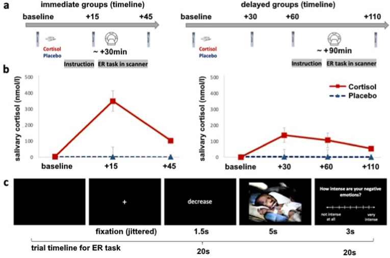
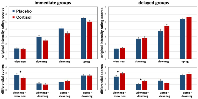
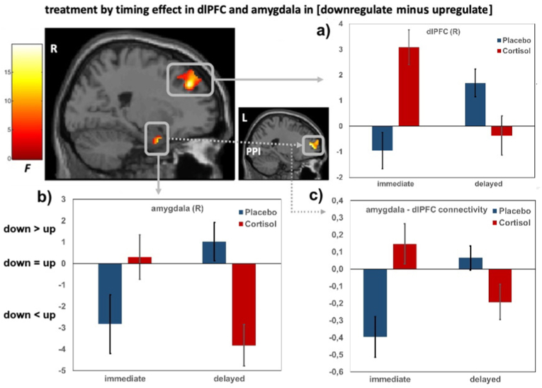
The ability of emotion regulation under stress is of crucial importance to psychosocial health. Yet, the dynamic function of stress hormones for the cognitive control of emotions over time via non-genomic and genomic cortisol effects remains to be elucidated. In this randomized, double-blind, placebo-controlled neuroimaging experiment, 105 participants (54 men, 51 women) received 20 mg hydrocortisone (cortisol) or a placebo either 30min (rapid, non-genomic cortisol effects) or 90min (slow, genomic cortisol effects) prior to a cognitive reappraisal task including different regulatory goals (i.e., downregulate vs. upregulate negative emotions). On the behavioral level, cortisol rapidly reduced and slowly enhanced emotional responsivity to negative pictures. However, only slow cortisol effects improved downregulation of negative emotions. On the neural level, cortisol rapidly enhanced, but slowly reduced amygdala and dorsolateral prefrontal activation as well as functional connectivity between both structures in the down- minus upregulate contrast. This interaction speaks for an effortful but ineffective regulation of negative emotions during rapid cortisol effects and improved emotion regulation capacities during slow cortisol effects. Taken together, these results indicate a functional shift of cortisol effects on emotion regulation processes over time which may foster successful adaptation to and recovery from stressful life events.

Keywords: Cognitive reappraisal; Functional magnetic resonance imaging; Genomic cortisol effects; Glucocorticoids; Stress hormones.


Conflict of interest statement

None.

Figures

Fig. 1
Fig. 1
Experimental design including the timing of cortisol/placebo administration with regard to the onset of the emotion regulation (ER) paradigm (a), cortisol concentrations over time (b) and an exemplary trial of the ER paradigm (c). a) The two immediate groups underwent the ER paradigm 25–30min after pharmacological manipulation. Participants of the two delayed groups were tested in the ER task 90–95min after tablet intake. Time points of saliva samples for cortisol concentrations slightly differed between the two datasets due to the different timing required for rapid vs. slow cortisol effects. b) In the line charts, mean cortisol concentrations (error bars represent standard errors of the mean) over time are illustrated separately for the cortisol and the placebo groups. Please note that peak cortisol concentrations of the two datasets were different, which reflect different sampling time points (15min (immediate group) and 30min (delayed group) after tablet intake) explaining the distinct result patterns. Of note, the observed cortisol concentrations after the intake of 20 mg hydrocortisone were elevated to supraphysiological levels. Thus, it is reasonable to assume that most mineralocorticoid and glucocorticoid receptors have been occupied in both datasets. Differences in these supraphysiological cortisol levels will most likely not be associated with any differential effect on ER processes. c) Trial timeline for the ER paradigm. The ER paradigm consisted of the following four conditions: view neutral, view negative, upregulate negative and downregulate negative. An instructional cue giving information about the condition was given before picture presentation (in this example, “decrease” for downregulate negative). Participants were required to rate the emotional intensity immediately after picture presentation.
Fig. 2
Fig. 2
Immediate and delayed cortisol effects on intensity ratings of the ER task. The left panel is from the immediate dataset including rapid cortisol effects, the right panel is from the delayed dataset including slow cortisol effects on intensity ratings. The top panel shows the original intensity ratings, the lower panel depicts difference scores (comparable to the fMRI contrasts). Opposing rapid and slow cortisol effects occurred for emotion induction (view neg – view neu). In addition, slow cortisol effects promote downregulation of negative emotions (view neg – downreg). Error bars represent standard errors of the mean, *p < .05. An alternative post hoc approach was realized with separate ANOVAs for each condition resulting in significant timing × treatment interactions in the view negative (F(1,97) = 9.28, p = .003, η2p = .09) and upregulate condition (F(1,97) = 4.94, p = .029, η2p = .05). When viewing negative pictures, intensity ratings were significantly lower in the cortisol relative to the placebo immediate group (F(1,46) = 4.58, p = .038, η2p = .09), but higher in the cortisol relative to the placebo delayed group (F(1,51) = 4.87, p = .032, η2p = .09). This pattern of results is consistent with our statistical approach reported above. For upregulate, post hoc tests conducted separately for the immediate and delayed group did not reveal any significant differences (all ps > .05). Separate analyses regarding the factor treatment revealed no differences between the immediate and delayed placebo group (p > .66), but higher intensity ratings for the delayed relative to the immediate cortisol group (F(1,47) = 7.75, p = .008, η2p = .14). For view neutral and downregulate, no significant main or interaction effects were observed.
Fig. 3
Fig. 3
Treatment by timing interaction in brain activation and functional connectivity in the downregulate minus upregulate contrast. a) cortisol administration induced a higher dlPFC activation in the immediate (i.e., immediate cortisol minus immediate placebo) compared to the delayed group (i.e., delayed cortisol – delayed placebo) in the contrast downregulate minus upregulate; b) cortisol (relative to placebo) increased amygdala activation in the immediate compared to the delayed cortisol group in the same contrast; c) cortisol (relative to placebo) increased amygdala-dlPFC functional connectivity in the immediate compared to the delayed group with downregulation as anchor of interpretation. Alternatively, cortisol (relative to placebo) induced more amygdala-dlPFC functional connectivity in the delayed group for upregulation. The average contrast estimates for significant peak voxels were extracted as bar graphs, with error bars representing standard errors of the mean. Please note that due to the arbitrary baseline, activations or deactivations cannot be inferred. But positive values reflect higher activations for downregulate relative to upregulate (down > up), negative values represent higher activations for upregulate compared to downregulate (down < up), values near zero reflect no activation differences between down- and upregulate (down = up).

Similar articles

Cited by

References

    1. Arnsten A.F.T. Stress signalling pathways that impair prefrontal cortex structure and function. Nat. Rev. Neurosci. 2009;10:410–422. doi: 10.1038/nrn2648. - DOI - PMC - PubMed
    1. Berboth S., Morawetz C. Amygdala-prefrontal connectivity during emotion regulation: a meta-analysis of psychophysiological interactions. Neuropsychologia. 2021;153 doi: 10.1016/j.neuropsychologia.2021.107767. - DOI - PubMed
    1. Buhle J.T., Silvers J.A., van Wager T.D., Lopez R., Onyemekwu C., Kober H., Weber J., Ochsner K.N. Cognitive reappraisal of emotion: a meta-analysis of human neuroimaging studies. Cerebr. Cortex. 2014;24:2981–2990. doi: 10.1093/cercor/bht154. - DOI - PMC - PubMed
    1. de Kloet E.R. Hormones and the stressed brain. Ann. N. Y. Acad. Sci. 2004;1018:1–15. doi: 10.1196/annals.1296.001. - DOI - PubMed
    1. de Kloet E.R., Karst H., Joëls M. Corticosteroid hormones in the central stress response: quick-and-slow. Front. Neuroendocrinol. 2008;29:268–272. doi: 10.1016/j.yfrne.2007.10.002. - DOI - PubMed

LinkOut - more resources