Case Reports
. 2023 May 21:49:102429.
doi: 10.1016/j.eucr.2023.102429. eCollection 2023 Jul.

Adrenal neuroblastoma in three year old boy, mistaken for pancreatic tumor: A case report

Affiliations
Case Reports

Adrenal neuroblastoma in three year old boy, mistaken for pancreatic tumor: A case report

Amina Karray et al. Urol Case Rep. .

Abstract

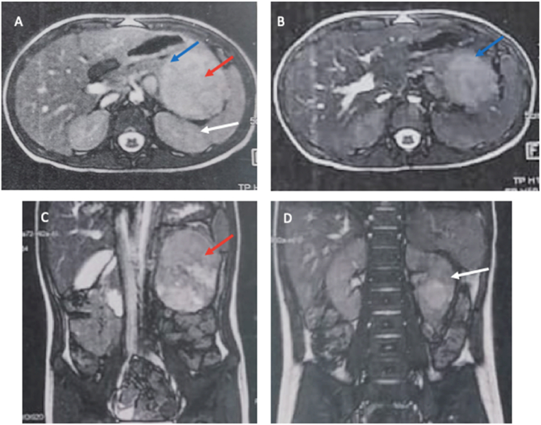
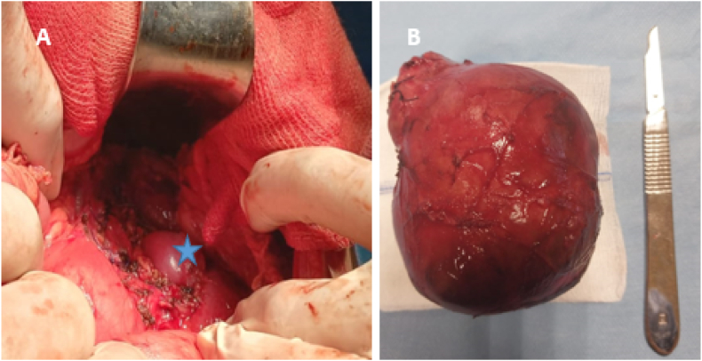
Neuroblastoma is the most common extracranial solid tumor in children, often manifests in the retroperitoneal region. We present a case of a 3-year-old boy with no previous medical history, presented for abdominal distension. Physical examination revealed a distinct, mobile, solid mass situated in the left lumbar region. Abdominal magnetic resonance imaging displayed a well delimited, well-encapsulated mass attached to the tail of the pancreas. Urinary catecholamine metabolite levels were negative. Surgical exploration revealed that the tumor was primitively related to the left adrenal gland, and a complete resection was performed. The postoperative recovery was uncomplicated. NMYC oncogene was non-amplified.

Keywords: Adrenal gland neoplasms; Neuroblastoma; Oncology; Pediatrics.


Conflict of interest statement

The authors declare no conflicts of interest regarding the publication of this article.

Figures

Fig. 1
Fig. 1
Abdominal MRI, (A–B) Axial T2 weighted images and (C–D) coronal T2 weighted images showing a large retro-peritoneal mass (red arrow): The mass is well encapsulated with heterogeneous high signal intensity. It deforms the edge of the tail of pancreas, in beak shape (blue arrow), suggestive that the mass originates from the pancreas. The left kidney (white arrowhead) is moderately displaced posteriorly. (For interpretation of the references to color in this figure legend, the reader is referred to the Web version of this article.)
Fig. 2
Fig. 2
(A) Tumoral bed after en-bloc resection with the left adrenal, leaving the left kidney in place (asterix). (B) Specimen.
Fig. 3
Fig. 3
(A) Macroscopic features of adrenal differentiating neuroblastic tumors. On gross section, the specimen is totally occupied by a largely hemorrhagic encapsulated tumor formation. The rest of the specimen is solid over 4 cm with a brownish color and some cystic formations. (B) Neuroblastoma (Schwannian stroma-poor), differentiating subtype (original, × 400). Microscopy revealed a tumor composed of small round blue cells forming rosettes with scant cytoplasm, Schwannian stroma-poor (arrow), hyperchromatic nuclei, and atypical mitoses. (For interpretation of the references to color in this figure legend, the reader is referred to the Web version of this article.)

References

    1. Matthay K., Maris J., Schleiermacher G., et al. Neuroblastoma. Nat Rev Dis Primers. 2016;2(2) - PubMed
    1. Croteau N., Nuchtern J., LaQuaglia M.P. Management of neuroblastoma in pediatric patients. Surg Oncol Clin. 2021;30(2):291–304. - PubMed
    1. Irwin M.S., Naranjo A., Zhang F.F., et al. Neuroblastoma risk classification system: a report from the children's Oncology Group. J Clin Oncol. 2021;39(29):3229–3241. - PMC - PubMed
    1. Monclair T., Brodeur G.M., Ambros P.F., et al. The international neuroblastoma risk Group (INRG) staging system: an INRG task force report. J Clin Oncol. 2009;27(2):298–303. - PMC - PubMed
    1. Lanza C., Galeazzi V., Carboni N., et al. Neuroblastoma image-defined risk factors in adrenal neuroblastoma: role of radiologist. Gland Surg. 2019;8(3):S168–S177. - PMC - PubMed

Publication types

LinkOut - more resources