Review
. 2023 May 18:14:1177670.
doi: 10.3389/fimmu.2023.1177670. eCollection 2023.

Lytic granule exocytosis at immune synapses: lessons from neuronal synapses

Affiliations
Review

Lytic granule exocytosis at immune synapses: lessons from neuronal synapses

Hsin-Fang Chang et al. Front Immunol. .

Abstract

Regulated exocytosis is a central mechanism of cellular communication. It is not only the basis for neurotransmission and hormone release, but also plays an important role in the immune system for the release of cytokines and cytotoxic molecules. In cytotoxic T lymphocytes (CTLs), the formation of the immunological synapse is required for the delivery of the cytotoxic substances such as granzymes and perforin, which are stored in lytic granules and released via exocytosis. The molecular mechanisms of their fusion with the plasma membrane are only partially understood. In this review, we discuss the molecular players involved in the regulated exocytosis of CTL, highlighting the parallels and differences to neuronal synaptic transmission. Additionally, we examine the strengths and weaknesses of both systems to study exocytosis.

Keywords: CD8+ cells; SNARE proteins; cytotoxic T lymphocytes; endocytosis; exocytosis; neuron; synapse.


Conflict of interest statement

The authors declare that the research was conducted in the absence of any commercial or financial relationships that could be construed as a potential conflict of interest.

Figures
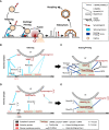
Figure 1
Figure 1
Model of the exocytosis machinery. (A) Schematic representation of the steps that SVs and LGs undergo before and after fusion with the plasma membrane. For sake of clarity only selected proteins involved in this process are shown. More comprehensive protein-protein interaction networks are shown in (BE). (B, C) Protein Interactions occurring during tethering (B) and docking/priming (C) of SVs in neurons. (D, E) Protein interaction required for tethering (D) and docking/priming (E) of LGs in CTL. Bottom legends applies to panels (B-E) The light green squares indicate the strong interaction of the t-SNAREs during tethering and of the entire SNARE complex during docking/priming. Lines indicate protein interactions, stippled lines show probable protein interactions. Black lines with blunt arrow correspond to inhibitory interactions.
Figure 2
Figure 2
CAPS2e, IA2 and snapin mRNA are found in mouse CD8+ T cells. (A) RT-PCR of murine naïve (d0) and day3 (d3) stimulated CTLs and spleen using primers specific for CAPS1 and all known CAPS2 splice variant as described in Nguyen Truong et al. (2014). Total RNA isolated from cerebellum was used as positive control, water was used as negative control and the housekeeping gene GAPDH as loading control. Note that only CAPS2e was detected in CTL and spleen. Data are representative of two independent experiments from two mice. (B, C) CTL mRNA expression profile of IA2 (B) and snapin (C) before and after 3 days of stimulation with anti-CD3/CD28 antibody-coated beads (d3). Unstimulated CTLs (d0) were harvested directly after CTLs isolation. Total RNA of adult mouse brain and kidney were used as positive control for snapin and IA2, respectively and H2O was used as the negative control for PCR. Data are representative of two independent experiments from two mice. See supplementary material file for materials and methods.

References

    1. Dustin ML. The immunological synapse. Cancer Immunol Res (2014) 2(11):1023–33. doi: 10.1158/2326-6066.Cir-14-0161 - DOI - PMC - PubMed
    1. Kaiser CA, Schekman R. Distinct sets of SEC genes govern transport vesicle formation and fusion early in the secretory pathway. Cell (1990) 61(4):723–33. doi: 10.1016/0092-8674(90)90483-u - DOI - PubMed
    1. Clary DO, Griff IC, Rothman JE. SNAPs, a family of NSF attachment proteins involved in intracellular membrane fusion in animals and yeast. Cell (1990) 61(4):709–21. doi: 10.1016/0092-8674(90)90482-t - DOI - PubMed
    1. Rothman JE, Orci L. Molecular dissection of the secretory pathway. Nature (1992) 355(6359):409–15. doi: 10.1038/355409a0 - DOI - PubMed
    1. Weber T, Zemelman BV, McNew JA, Westermann B, Gmachl M, Parlati F, et al. . SNAREpins: minimal machinery for membrane fusion. Cell (1998) 92(6):759–72. doi: 10.1016/S0092-8674(00)81404-X - DOI - PubMed

Publication types